Figure 3
Epstein-Barr infection causing toxic epidermal necrolysis, hemophagocytic lymphohistiocytosis and cerebritis in a pediatric patient
Aikaterini Solomou*, Vasileios Patriarcheas, Pantelis Kraniotis and Andreas Eliades
Published: 18 March, 2020 | Volume 4 - Issue 1 | Pages: 015-019
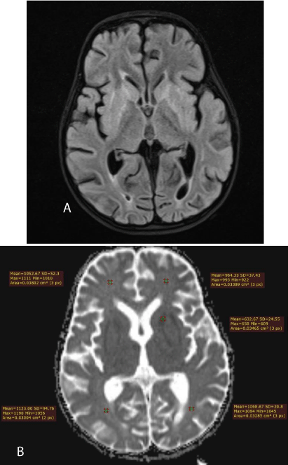
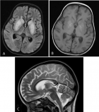
Figure 3:
a) 2nd MRI. The high signal intensity mainly affects the internal and external capsules. b) 2nd MRI. Axial ADC map. There is true diffusion restriction only in the external and internal capsules, according the low ADC values, as measured.
Read Full Article HTML DOI: 10.29328/journal.acr.1001032 Cite this Article Read Full Article PDF
More Images
Similar Articles
-
Trichomonas Vaginalis-A Clinical ImageAstrit M Gashi*. Trichomonas Vaginalis-A Clinical Image. . 2017 doi: 10.29328/journal.hjcr.1001001; 1: 001-002
-
Gastric Mucosal CalcinosisVedat Goral*,Irem Ozover,Ilknur Turkmen. Gastric Mucosal Calcinosis. . 2017 doi: 10.29328/journal.hjcr.1001002; 1: 003-005
-
Giant Lipoma Anterior Neck: A case reportManu CB*,Gowri Sankar M,Arun Alexander. Giant Lipoma Anterior Neck: A case report . . 2017 doi: 10.29328/journal.hjcr.1001003; 1: 006-008
-
Catamenial pneumothorax: Presentation of an uncommon PathologyRui Haddad*,Caterin Arévalo,David Nigri. Catamenial pneumothorax: Presentation of an uncommon Pathology . . 2017 doi: 10.29328/journal.hjcr.1001004; 1: 009-013
-
A rare case: Congenital Megalourethra in prune belly syndromeNuman Baydilli*,Ismail Selvi,Emre Can Akınsal. A rare case: Congenital Megalourethra in prune belly syndrome. . 2018 doi: 10.29328/journal.acr.1001005; 2: 001-003
-
Trauma to the neck: Manifestation of injuries outside the original zone of injury-A case reportStephen O Heard*,Alexander Christakis,Brian Tashjian,Anne M Gilroy3. Trauma to the neck: Manifestation of injuries outside the original zone of injury-A case report. . 2018 doi: 10.29328/journal.acr.1001006; 2: 004-009
-
Meige Trofoedema: A form of primary lymphedemaCarlos Al Sanchez Salguero*. Meige Trofoedema: A form of primary lymphedema. . 2018 doi: 10.29328/journal.acr.1001007; 2: 010-014
-
A rare case of Diabetic Foot in male of middle age has been shown Diabetic footSujit K Bhattacharya*. A rare case of Diabetic Foot in male of middle age has been shown Diabetic foot. . 2018 doi: 10.29328/journal.acr.1001008; 2: 015-015
-
Brooke-Spiegler Syndrome: A rare cause of skin appendageal tumorN Suganthan*,S Pirasath,DD Dikowita. Brooke-Spiegler Syndrome: A rare cause of skin appendageal tumor. . 2018 doi: 10.29328/journal.acr.1001009; 2: 016-018
-
McArdle’s Disease (Glycogen Storage Disease type V): A Clinical CaseCameselle-Teijeiro JF*,Calheiros-Cruz T,Caamaño-Vara MP,Villar-Fernández B,Ruibal-Azevedo J,Cameselle-Cortizo L,Cameselle-Arias M,Charro Gamallo ME,Turienzo-Pacho F,Yera Acosta A. McArdle’s Disease (Glycogen Storage Disease type V): A Clinical Case. . 2018 doi: 10.29328/journal.acr.1001010; 2: 019-023
Recently Viewed
-
Navigating Weight Management with Stevia: Insights into Glycemic ControlShashikanth Kharat*, Sanjana Mali*. Navigating Weight Management with Stevia: Insights into Glycemic Control. New Insights Obes Gene Beyond. 2024: doi: 10.29328/journal.niogb.1001021; 8: 006-008
-
Microbial Conversion and Utilization of CO2Ge-Ge Wang, Yuan Zhang, Xiao-Yan Wang*, Gen-Lin Zhang. Microbial Conversion and Utilization of CO2. Ann Civil Environ Eng. 2023: doi: 10.29328/journal.acee.1001055; 7: 045-060
-
Role of RBC Parameters to Differentiate between Iron Deficiency Anemia and Anemia of Chronic DiseasesReena Bhaisare*, Ravindranath M, Gurmeet Singh. Role of RBC Parameters to Differentiate between Iron Deficiency Anemia and Anemia of Chronic Diseases. J Hematol Clin Res. 2023: doi: 10.29328/journal.jhcr.1001024; 7: 021-024
-
Causes of hospital admission of chronic kidney disease patient in a tertiary kidney care hospitalSalman Imtiaz*,Ruqaya Qureshi,Amna Hamid,Beena Salman,Murtaza F Drohlia,Aasim Ahmad. Causes of hospital admission of chronic kidney disease patient in a tertiary kidney care hospital. J Clini Nephrol. 2019: doi: 10.29328/journal.jcn.1001033; 3: 100-106
-
The inflammatory profile of chronic kidney disease patientsHanen Chaker,Faiçal Jarraya,Salma Toumi*,Khawla Kammoun,Hichem Mahfoudh,Fatma Ayadi,Soumaya Yaich,Mohamed Ben Hmida. The inflammatory profile of chronic kidney disease patients. J Clini Nephrol. 2021: doi: 10.29328/journal.jcn.1001083; 5: 107-111
Most Viewed
-
Evaluation of Biostimulants Based on Recovered Protein Hydrolysates from Animal By-products as Plant Growth EnhancersH Pérez-Aguilar*, M Lacruz-Asaro, F Arán-Ais. Evaluation of Biostimulants Based on Recovered Protein Hydrolysates from Animal By-products as Plant Growth Enhancers. J Plant Sci Phytopathol. 2023 doi: 10.29328/journal.jpsp.1001104; 7: 042-047
-
Feasibility study of magnetic sensing for detecting single-neuron action potentialsDenis Tonini,Kai Wu,Renata Saha,Jian-Ping Wang*. Feasibility study of magnetic sensing for detecting single-neuron action potentials. Ann Biomed Sci Eng. 2022 doi: 10.29328/journal.abse.1001018; 6: 019-029
-
Physical activity can change the physiological and psychological circumstances during COVID-19 pandemic: A narrative reviewKhashayar Maroufi*. Physical activity can change the physiological and psychological circumstances during COVID-19 pandemic: A narrative review. J Sports Med Ther. 2021 doi: 10.29328/journal.jsmt.1001051; 6: 001-007
-
Pediatric Dysgerminoma: Unveiling a Rare Ovarian TumorFaten Limaiem*, Khalil Saffar, Ahmed Halouani. Pediatric Dysgerminoma: Unveiling a Rare Ovarian Tumor. Arch Case Rep. 2024 doi: 10.29328/journal.acr.1001087; 8: 010-013
-
Prospective Coronavirus Liver Effects: Available KnowledgeAvishek Mandal*. Prospective Coronavirus Liver Effects: Available Knowledge. Ann Clin Gastroenterol Hepatol. 2023 doi: 10.29328/journal.acgh.1001039; 7: 001-010

HSPI: We're glad you're here. Please click "create a new Query" if you are a new visitor to our website and need further information from us.
If you are already a member of our network and need to keep track of any developments regarding a question you have already submitted, click "take me to my Query."


