Figure 4
Epstein-Barr infection causing toxic epidermal necrolysis, hemophagocytic lymphohistiocytosis and cerebritis in a pediatric patient
Aikaterini Solomou*, Vasileios Patriarcheas, Pantelis Kraniotis and Andreas Eliades
Published: 18 March, 2020 | Volume 4 - Issue 1 | Pages: 015-019
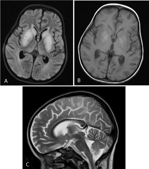
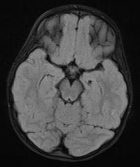
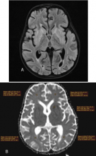
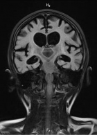
Figure 4:
a) 3rd MRI. Axial FLAIR. There is diffuse hyperintensity in the basal ganglia and caudate nuclei. b) 3rd MRI. Axial T1WI. Hyperintensity in the putamen and globus pallidus bilaterally may represent methemoglobin, due to petechial hemorrhage. c) 3rd MRI SAG T2WI. The corpus callosum is slightly thinned with abnormal signal in its posterior part.
Read Full Article HTML DOI: 10.29328/journal.acr.1001032 Cite this Article Read Full Article PDF
More Images
Similar Articles
-
Trichomonas Vaginalis-A Clinical ImageAstrit M Gashi*. Trichomonas Vaginalis-A Clinical Image. . 2017 doi: 10.29328/journal.hjcr.1001001; 1: 001-002
-
Gastric Mucosal CalcinosisVedat Goral*,Irem Ozover,Ilknur Turkmen. Gastric Mucosal Calcinosis. . 2017 doi: 10.29328/journal.hjcr.1001002; 1: 003-005
-
Giant Lipoma Anterior Neck: A case reportManu CB*,Gowri Sankar M,Arun Alexander. Giant Lipoma Anterior Neck: A case report . . 2017 doi: 10.29328/journal.hjcr.1001003; 1: 006-008
-
Catamenial pneumothorax: Presentation of an uncommon PathologyRui Haddad*,Caterin Arévalo,David Nigri. Catamenial pneumothorax: Presentation of an uncommon Pathology . . 2017 doi: 10.29328/journal.hjcr.1001004; 1: 009-013
-
A rare case: Congenital Megalourethra in prune belly syndromeNuman Baydilli*,Ismail Selvi,Emre Can Akınsal. A rare case: Congenital Megalourethra in prune belly syndrome. . 2018 doi: 10.29328/journal.acr.1001005; 2: 001-003
-
Trauma to the neck: Manifestation of injuries outside the original zone of injury-A case reportStephen O Heard*,Alexander Christakis,Brian Tashjian,Anne M Gilroy3. Trauma to the neck: Manifestation of injuries outside the original zone of injury-A case report. . 2018 doi: 10.29328/journal.acr.1001006; 2: 004-009
-
Meige Trofoedema: A form of primary lymphedemaCarlos Al Sanchez Salguero*. Meige Trofoedema: A form of primary lymphedema. . 2018 doi: 10.29328/journal.acr.1001007; 2: 010-014
-
A rare case of Diabetic Foot in male of middle age has been shown Diabetic footSujit K Bhattacharya*. A rare case of Diabetic Foot in male of middle age has been shown Diabetic foot. . 2018 doi: 10.29328/journal.acr.1001008; 2: 015-015
-
Brooke-Spiegler Syndrome: A rare cause of skin appendageal tumorN Suganthan*,S Pirasath,DD Dikowita. Brooke-Spiegler Syndrome: A rare cause of skin appendageal tumor. . 2018 doi: 10.29328/journal.acr.1001009; 2: 016-018
-
McArdle’s Disease (Glycogen Storage Disease type V): A Clinical CaseCameselle-Teijeiro JF*,Calheiros-Cruz T,Caamaño-Vara MP,Villar-Fernández B,Ruibal-Azevedo J,Cameselle-Cortizo L,Cameselle-Arias M,Charro Gamallo ME,Turienzo-Pacho F,Yera Acosta A. McArdle’s Disease (Glycogen Storage Disease type V): A Clinical Case. . 2018 doi: 10.29328/journal.acr.1001010; 2: 019-023
Recently Viewed
-
Correlation between the Values of Immature Platelet Fraction and Mean Platelet Volume with the Extent of Coronary Artery Disease in Patients with Non-ST-Segment Elevation Myocardial InfarctionShadab Rauf*, Tarun Kumar, Vijay Kumar, Ranjit Kumar Nath. Correlation between the Values of Immature Platelet Fraction and Mean Platelet Volume with the Extent of Coronary Artery Disease in Patients with Non-ST-Segment Elevation Myocardial Infarction. J Cardiol Cardiovasc Med. 2023: doi: 10.29328/journal.jccm.1001163; 8: 114-121
-
A Water-soluble Form of Dihydroquercetin Reduces LPS-induced Astrogliosis, Vascular Remodeling, and mRNA VEGF-A Levels in the Substantia Nigra of Aged RatsElena S Alalykina, Tatyana N Sergeeva, Michail A Ananyan*, Ivan A Cherenkov, Valeriy G Sergeev. A Water-soluble Form of Dihydroquercetin Reduces LPS-induced Astrogliosis, Vascular Remodeling, and mRNA VEGF-A Levels in the Substantia Nigra of Aged Rats. J Neurosci Neurol Disord. 2024: doi: 10.29328/journal.jnnd.1001092; 8: 014-019
-
Relationship between the Level of Spirituality and Blood Pressure Control among Adult Hypertensive Patients in a Southwestern Community in NigeriaAdetunji OMONIJO*, Paul OLOWOYO, Azeez Oyemomi IBRAHIM, Segun Matthew AGBOOLA, Oluwaserimi Adewumi AJETUNMOBI, Temitope Moronkeji OLANREWAJU, Adejumoke Oluwatosin OMONIJO. Relationship between the Level of Spirituality and Blood Pressure Control among Adult Hypertensive Patients in a Southwestern Community in Nigeria. Ann Clin Hypertens. 2023: doi: 10.29328/journal.ach.1001034; 7: 004-012
-
Morning and Evening Exercise with or without High-Pressure Intervals have Different Effects on Controlling Blood Sugar and Glucose Fluctuations in People with Type 1 and Type 2 DiabetesReza Sheikh*. Morning and Evening Exercise with or without High-Pressure Intervals have Different Effects on Controlling Blood Sugar and Glucose Fluctuations in People with Type 1 and Type 2 Diabetes . Int J Bone Marrow Res. 2023: doi: 10.29328/journal.ijbmr.1001016; 6: 001-003
-
Assessment of the Quality of Life of the Caregiver of Pediatric Patients with Chronic Kidney Disease in a Tertiary Health Care FacilityAsma Hamoud Alruwaili*, Khaled Abdulaziz Alsaran and Abdulaziz Abdullah Alshathri. Assessment of the Quality of Life of the Caregiver of Pediatric Patients with Chronic Kidney Disease in a Tertiary Health Care Facility. J Clini Nephrol. 2024: doi: 10.29328/journal.jcn.1001123; 8: 032-038
Most Viewed
-
Evaluation of Biostimulants Based on Recovered Protein Hydrolysates from Animal By-products as Plant Growth EnhancersH Pérez-Aguilar*, M Lacruz-Asaro, F Arán-Ais. Evaluation of Biostimulants Based on Recovered Protein Hydrolysates from Animal By-products as Plant Growth Enhancers. J Plant Sci Phytopathol. 2023 doi: 10.29328/journal.jpsp.1001104; 7: 042-047
-
Feasibility study of magnetic sensing for detecting single-neuron action potentialsDenis Tonini,Kai Wu,Renata Saha,Jian-Ping Wang*. Feasibility study of magnetic sensing for detecting single-neuron action potentials. Ann Biomed Sci Eng. 2022 doi: 10.29328/journal.abse.1001018; 6: 019-029
-
Physical activity can change the physiological and psychological circumstances during COVID-19 pandemic: A narrative reviewKhashayar Maroufi*. Physical activity can change the physiological and psychological circumstances during COVID-19 pandemic: A narrative review. J Sports Med Ther. 2021 doi: 10.29328/journal.jsmt.1001051; 6: 001-007
-
Pediatric Dysgerminoma: Unveiling a Rare Ovarian TumorFaten Limaiem*, Khalil Saffar, Ahmed Halouani. Pediatric Dysgerminoma: Unveiling a Rare Ovarian Tumor. Arch Case Rep. 2024 doi: 10.29328/journal.acr.1001087; 8: 010-013
-
Prospective Coronavirus Liver Effects: Available KnowledgeAvishek Mandal*. Prospective Coronavirus Liver Effects: Available Knowledge. Ann Clin Gastroenterol Hepatol. 2023 doi: 10.29328/journal.acgh.1001039; 7: 001-010

HSPI: We're glad you're here. Please click "create a new Query" if you are a new visitor to our website and need further information from us.
If you are already a member of our network and need to keep track of any developments regarding a question you have already submitted, click "take me to my Query."


