Figure 2
Pediatric Dysgerminoma: Unveiling a Rare Ovarian Tumor
Faten Limaiem*, Khalil Saffar and Ahmed Halouani
Published: 19 January, 2024 | Volume 8 - Issue 1 | Pages: 010-013

Figure 2:
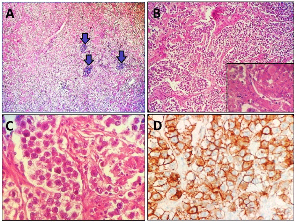
Figure 2: A: Tumor proliferation arranged in trabeculae and sheets separated by fibrous septa containing lymphoid aggregates (arrows) (Hematoxylin and eosin, magnification × 40). B: Tumor proliferation arranged in trabeculae and sheets separated by fibrous septa (Hematoxylin and eosin, magnification × 100). Inset: Presence of a granuloma in the tumor proliferation (Hematoxylin and eosin, magnification × 400). C: Higher power image showing polygonal cells with distinct cell borders and eosinophilic to clear cytoplasm. The nuclei are round and vesicular with prominent nucleoli. (Hematoxylin and eosin, magnification × 400).
D: Immunohistochemical study showed positive immunostaining of tumor cells with CD117. (Immunohistochemistry, magnification × 400).
Read Full Article HTML DOI: 10.29328/journal.acr.1001087 Cite this Article Read Full Article PDF
More Images
Similar Articles
-
Gastric Mucosal CalcinosisVedat Goral*,Irem Ozover,Ilknur Turkmen. Gastric Mucosal Calcinosis. . 2017 doi: 10.29328/journal.hjcr.1001002; 1: 003-005
-
Catamenial pneumothorax: Presentation of an uncommon PathologyRui Haddad*,Caterin Arévalo,David Nigri. Catamenial pneumothorax: Presentation of an uncommon Pathology . . 2017 doi: 10.29328/journal.hjcr.1001004; 1: 009-013
-
Treatment of autoimmune hemolytic anemia with erythropoietin: A case reportRenato A Guzmán*,Juan P Ovalle,Estefanía M Orozco,Laura C Pedraza,María C Barrera,Dormar D Barrios M. Treatment of autoimmune hemolytic anemia with erythropoietin: A case report. . 2019 doi: 10.29328/journal.acr.1001022; 3: 043-046
-
Foley catheter balloon tamponade as a method of controlling iatrogenic pulmonary artery bleeding in redo thoracic surgeryMarcus Taylor*,Muhammad Asghar Nawaz,Ozhin Karadakhy,Denish Apparau,Kandadai Rammohan,Paul Waterworth. Foley catheter balloon tamponade as a method of controlling iatrogenic pulmonary artery bleeding in redo thoracic surgery. . 2019 doi: 10.29328/journal.acr.1001023; 3: 047-049
-
Scraping cytology and scanning electron microscopy in diagnosis and therapy of corneal ulcer by mycobacterium infectionRusso Giacomo*,Del Prete Salvatore,Del Prete Antonio,Meloni Marisa,Capaldi Roberto,Grumetto Lucia. Scraping cytology and scanning electron microscopy in diagnosis and therapy of corneal ulcer by mycobacterium infection. . 2019 doi: 10.29328/journal.acr.1001024; 3: 050-053
-
Orgasmic coitus triggered stillbirth via placental abruption: A case reportAttila Pajor*,Semmelweis University Faculty of Medicine, Department of Obstetrics and Gynecology, Budapest, Hungary,Márton Vezér,Henriette Pusztafalvi,Bianka Pencz,Semmelweis University II. Department of Pathology, Budapest, Hungary . Orgasmic coitus triggered stillbirth via placental abruption: A case report. . 2019 doi: 10.29328/journal.acr.1001026; 3: 056-058
-
A case study on Erdheim ‐ Chester DiseaseHarald Koeck*,Jakob Erdheim. A case study on Erdheim ‐ Chester Disease. . 2020 doi: 10.29328/journal.acr.1001027; 4: 001-003
-
Acute and post burn reconstructive surgery of the female trunk with artificial dermis to facilitate healthy pregnancyDantzer E*,Campech G,Mazanovich M. Acute and post burn reconstructive surgery of the female trunk with artificial dermis to facilitate healthy pregnancy. . 2020 doi: 10.29328/journal.acr.1001036; 4: 028-031
-
Chronic subdural haematoma associated with arachnoid cyst of the middle fossa in a soccer player: Case report and review of the literatureElena Beretta*,Michele Incerti,Giuseppe Raudino,Gaspare F Montemagno,Franco Servadei. Chronic subdural haematoma associated with arachnoid cyst of the middle fossa in a soccer player: Case report and review of the literature. . 2020 doi: 10.29328/journal.acr.1001037; 4: 032-037
-
Exceptional intraoperative aspects of mesenteric venous gasWael Ferjaoui*,Mohamed Hajri,Aziz Atallah,Rached Bayar,Dhouha Bacha,Mohamed Tahar Khalfallah. Exceptional intraoperative aspects of mesenteric venous gas. . 2020 doi: 10.29328/journal.acr.1001042; 4: 050-051
Recently Viewed
-
Bladder Benign Inverted Papilloma in Young Men: A Case ReportMehdi Marrak*, Yassine Ouanes, Kays Chaker, Moez Rahoui, Mokhtar Bibi, Yassine Nouira. Bladder Benign Inverted Papilloma in Young Men: A Case Report. Arch Case Rep. 2024: doi: 10.29328/journal.acr.1001093; 8: 048-049
-
Impact of Chronic Kidney Disease on Major Adverse Cardiac Events in Patients with Acute Myocardial Infarction: A Retrospective Cohort StudyAbbas Andishmand, Ehsan Zolfeqari*, Mahdiah Sadat Namayandah, Hossein Montazer Ghaem. Impact of Chronic Kidney Disease on Major Adverse Cardiac Events in Patients with Acute Myocardial Infarction: A Retrospective Cohort Study. J Cardiol Cardiovasc Med. 2024: doi: 10.29328/journal.jccm.1001175; 9: 029-034
-
Persistent Bilateral Vocal Cord Paralysis Following Unilateral Basal Ganglia HemorrhageNikil Joseph John, John Thomas*, Martyn Bracewell. Persistent Bilateral Vocal Cord Paralysis Following Unilateral Basal Ganglia Hemorrhage. J Cardiol Cardiovasc Med. 2024: doi: 10.29328/journal.jccm.1001174; 9: 026-028.
-
The Effect of SGLT-2i and GLP-1RA on Major Cardiovascular Conditions: A Meta-AnalysisArjun V Jogimahanti*, Kevin A Honan, Talha Ahmed, Luis Leon-Novelo, Tarif Khair. The Effect of SGLT-2i and GLP-1RA on Major Cardiovascular Conditions: A Meta-Analysis. J Cardiol Cardiovasc Med. 2024: doi: 10.29328/journal.jccm.1001173; 9: 014-025
-
Aspirin for Primary Prevention of Cardiovascular Disease: What We Now KnowSteven M Weisman*,Dominick J Angiolillo. Aspirin for Primary Prevention of Cardiovascular Disease: What We Now Know. J Cardiol Cardiovasc Med. 2024: doi: 10.29328/journal.jccm.1001172; 9: 006-013
Most Viewed
-
Evaluation of Biostimulants Based on Recovered Protein Hydrolysates from Animal By-products as Plant Growth EnhancersH Pérez-Aguilar*, M Lacruz-Asaro, F Arán-Ais. Evaluation of Biostimulants Based on Recovered Protein Hydrolysates from Animal By-products as Plant Growth Enhancers. J Plant Sci Phytopathol. 2023 doi: 10.29328/journal.jpsp.1001104; 7: 042-047
-
Feasibility study of magnetic sensing for detecting single-neuron action potentialsDenis Tonini,Kai Wu,Renata Saha,Jian-Ping Wang*. Feasibility study of magnetic sensing for detecting single-neuron action potentials. Ann Biomed Sci Eng. 2022 doi: 10.29328/journal.abse.1001018; 6: 019-029
-
Physical activity can change the physiological and psychological circumstances during COVID-19 pandemic: A narrative reviewKhashayar Maroufi*. Physical activity can change the physiological and psychological circumstances during COVID-19 pandemic: A narrative review. J Sports Med Ther. 2021 doi: 10.29328/journal.jsmt.1001051; 6: 001-007
-
Pediatric Dysgerminoma: Unveiling a Rare Ovarian TumorFaten Limaiem*, Khalil Saffar, Ahmed Halouani. Pediatric Dysgerminoma: Unveiling a Rare Ovarian Tumor. Arch Case Rep. 2024 doi: 10.29328/journal.acr.1001087; 8: 010-013
-
Prospective Coronavirus Liver Effects: Available KnowledgeAvishek Mandal*. Prospective Coronavirus Liver Effects: Available Knowledge. Ann Clin Gastroenterol Hepatol. 2023 doi: 10.29328/journal.acgh.1001039; 7: 001-010

HSPI: We're glad you're here. Please click "create a new Query" if you are a new visitor to our website and need further information from us.
If you are already a member of our network and need to keep track of any developments regarding a question you have already submitted, click "take me to my Query."


-zohair-moh’d-allouh.jpg)


