Figure 2
Imaging aspect of neuromyelitis optica: a case report and review of the literature
H Lhajoui*, I Bounnit, N Moussali, A Merzem, O Amriss, H Belgadir and N Elbenna
Published: 07 December, 2021 | Volume 5 - Issue 2 | Pages: 036-038

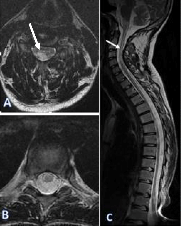
Figure 2:
Spinal Cord MRI on a axial (A,B), sagittal (C), showed signal hyper-intensities at cervical and thoracic spinal cord on the T2 weighted sequence, spans over 3 contiguous vertebral.
Read Full Article HTML DOI: 10.29328/journal.acr.1001055 Cite this Article Read Full Article PDF
More Images
Similar Articles
-
Imaging aspect of neuromyelitis optica: a case report and review of the literatureH Lhajoui*,I Bounnit,N Moussali,A Merzem,O Amriss,H Belgadir,N Elbenna. Imaging aspect of neuromyelitis optica: a case report and review of the literature. . 2021 doi: 10.29328/journal.acr.1001055; 5: 036-038
-
Influence of corneal spherical aberration, anterior chamber depth, and ocular axial length on the visual outcome with an extended depth of focus wavefront-designed intraocular lensBedei Andrea*,Carbonara Claudio,Farcomeni Alessio,Castellini Laura,Pietrelli Alessia. Influence of corneal spherical aberration, anterior chamber depth, and ocular axial length on the visual outcome with an extended depth of focus wavefront-designed intraocular lens. . 2022 doi: 10.29328/journal.acr.1001061; 6: 017-021
Recently Viewed
-
Fifteen year Follow-up of Relapsed/ Refractory Patients with Hodgkin Lymphoma Treated with Autologous Hematopoietic Stem Cell TransplantationSabree Abredabo*, Harrah Chiang, Leonard Klein, Tulio Rodriguez, Jacob D Bitran. Fifteen year Follow-up of Relapsed/ Refractory Patients with Hodgkin Lymphoma Treated with Autologous Hematopoietic Stem Cell Transplantation. J Stem Cell Ther Transplant. 2024: doi: 10.29328/journal.jsctt.1001040; 8: 033-037
-
Beta-1 Receptor (β1) in the Heart Specific Indicate to StereoselectivityRezk Rezk Ayyad*, Ahmed Mohamed Mansour, Ahmed Mohamed Nejm, Yasser Abdel Allem Hassan, Norhan Hassan Gabr, Ahmed Rezk Ayyad. Beta-1 Receptor (β1) in the Heart Specific Indicate to Stereoselectivity. Arch Pharm Pharma Sci. 2024: doi: 10.29328/journal.apps.1001060; 8: 082-088
-
Schizoaffective Disorder in an Individual with Mowat-Wilson Syndrome (MWS)Yadwinder Chuhan, Nimrit Bath, Muhammad Ayub*. Schizoaffective Disorder in an Individual with Mowat-Wilson Syndrome (MWS). Arch Psychiatr Ment Health. 2024: doi: 10.29328/journal.apmh.1001050; 8: 008-011
-
Mapping the Psychosocial: Introducing a Standardised System to Improve Psychosocial Understanding within Mental HealthMatthew Bretton Oakes*. Mapping the Psychosocial: Introducing a Standardised System to Improve Psychosocial Understanding within Mental Health. Arch Psychiatr Ment Health. 2024: doi: 10.29328/journal.apmh.1001051; 8: 012-019
-
Approaching Mental Health Through a Preventive Data Analysis PlatformGabriel F Pestana, Olga Valentim*. Approaching Mental Health Through a Preventive Data Analysis Platform. Arch Psychiatr Ment Health. 2024: doi: 10.29328/journal.apmh.1001052; 8: 020-027
Most Viewed
-
Evaluation of Biostimulants Based on Recovered Protein Hydrolysates from Animal By-products as Plant Growth EnhancersH Pérez-Aguilar*, M Lacruz-Asaro, F Arán-Ais. Evaluation of Biostimulants Based on Recovered Protein Hydrolysates from Animal By-products as Plant Growth Enhancers. J Plant Sci Phytopathol. 2023 doi: 10.29328/journal.jpsp.1001104; 7: 042-047
-
Feasibility study of magnetic sensing for detecting single-neuron action potentialsDenis Tonini,Kai Wu,Renata Saha,Jian-Ping Wang*. Feasibility study of magnetic sensing for detecting single-neuron action potentials. Ann Biomed Sci Eng. 2022 doi: 10.29328/journal.abse.1001018; 6: 019-029
-
Physical activity can change the physiological and psychological circumstances during COVID-19 pandemic: A narrative reviewKhashayar Maroufi*. Physical activity can change the physiological and psychological circumstances during COVID-19 pandemic: A narrative review. J Sports Med Ther. 2021 doi: 10.29328/journal.jsmt.1001051; 6: 001-007
-
Evaluation of In vitro and Ex vivo Models for Studying the Effectiveness of Vaginal Drug Systems in Controlling Microbe Infections: A Systematic ReviewMohammad Hossein Karami*, Majid Abdouss*, Mandana Karami. Evaluation of In vitro and Ex vivo Models for Studying the Effectiveness of Vaginal Drug Systems in Controlling Microbe Infections: A Systematic Review. Clin J Obstet Gynecol. 2023 doi: 10.29328/journal.cjog.1001151; 6: 201-215
-
Pediatric Dysgerminoma: Unveiling a Rare Ovarian TumorFaten Limaiem*, Khalil Saffar, Ahmed Halouani. Pediatric Dysgerminoma: Unveiling a Rare Ovarian Tumor. Arch Case Rep. 2024 doi: 10.29328/journal.acr.1001087; 8: 010-013

HSPI: We're glad you're here. Please click "create a new Query" if you are a new visitor to our website and need further information from us.
If you are already a member of our network and need to keep track of any developments regarding a question you have already submitted, click "take me to my Query."


