Figure 2
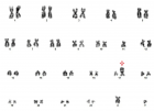
An unusual clonal chromosome abnormality der(17)t(11;17)(q24;p13)inv(11)(q13;q23) in a patient with chronic lymphocytic leukemia
Valentina Monti*, Fabio Serpenti, Lucia Farina, Maria Luisa Moiraghi, Maria Adele Testi and Giancarlo Pruneri
Published: 10 November, 2021 | Volume 5 - Issue 2 | Pages: 028-031
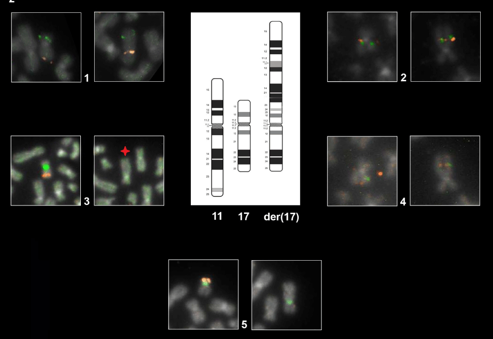
Figure 2:
Representative FISH Panel probes. The first eight cells each with two boxes marked with the number 1-2-3-4 show the normal ch.11 (left box) and the der(17) (right box) hybridized with different probes as follow: Box 1: WT1/FLI1 by combination probe set. FISH result highlight the der(17) orientation: green signal (WT1) map on the short arm of chromosome 11 and orange signal (FLI1) map on the long arm of chromosome 11. Box 2: CCND1 Dual Color Break Apart Probe Rearrangement. Der(17) harboring the CCND1 gene in closed proximity to 17p compared with normal chromosome. Box 3 LSI ATM/CEP11. As shown on normal ch.11 (left box) the orange fluorochrome hybridizes ATM gene and the green fluorochrome direct labeled the CEP11. The star on der(17) (right box) show the interstitial deletion affecting the gene ATM and the loss of CEP11. Box 4: MLL Dual Color Break Apart rearrangement. Der(17) harboring the MLL gene in closed proximity to 11p. The last two boxes, marked with the number 5, shows the normal ch.17 (left box) and der(17) in the (right box) hybridized with TP53 (orange)/CEP17(green). As illustrated der(17) keeps intact the CEP17.
Read Full Article HTML DOI: 10.29328/journal.acr.1001053 Cite this Article Read Full Article PDF
More Images
Similar Articles
-
An unusual clonal chromosome abnormality der(17)t(11;17)(q24;p13)inv(11)(q13;q23) in a patient with chronic lymphocytic leukemiaValentina Monti*,Fabio Serpenti,Lucia Farina,Maria Luisa Moiraghi,Maria Adele Testi,Giancarlo Pruneri. An unusual clonal chromosome abnormality der(17)t(11;17)(q24;p13)inv(11)(q13;q23) in a patient with chronic lymphocytic leukemia. . 2021 doi: 10.29328/journal.acr.1001053; 5: 028-031
Recently Viewed
-
Fifteen year Follow-up of Relapsed/ Refractory Patients with Hodgkin Lymphoma Treated with Autologous Hematopoietic Stem Cell TransplantationSabree Abredabo*, Harrah Chiang, Leonard Klein, Tulio Rodriguez, Jacob D Bitran. Fifteen year Follow-up of Relapsed/ Refractory Patients with Hodgkin Lymphoma Treated with Autologous Hematopoietic Stem Cell Transplantation. J Stem Cell Ther Transplant. 2024: doi: 10.29328/journal.jsctt.1001040; 8: 033-037
-
Beta-1 Receptor (β1) in the Heart Specific Indicate to StereoselectivityRezk Rezk Ayyad*, Ahmed Mohamed Mansour, Ahmed Mohamed Nejm, Yasser Abdel Allem Hassan, Norhan Hassan Gabr, Ahmed Rezk Ayyad. Beta-1 Receptor (β1) in the Heart Specific Indicate to Stereoselectivity. Arch Pharm Pharma Sci. 2024: doi: 10.29328/journal.apps.1001060; 8: 082-088
-
Schizoaffective Disorder in an Individual with Mowat-Wilson Syndrome (MWS)Yadwinder Chuhan, Nimrit Bath, Muhammad Ayub*. Schizoaffective Disorder in an Individual with Mowat-Wilson Syndrome (MWS). Arch Psychiatr Ment Health. 2024: doi: 10.29328/journal.apmh.1001050; 8: 008-011
-
Mapping the Psychosocial: Introducing a Standardised System to Improve Psychosocial Understanding within Mental HealthMatthew Bretton Oakes*. Mapping the Psychosocial: Introducing a Standardised System to Improve Psychosocial Understanding within Mental Health. Arch Psychiatr Ment Health. 2024: doi: 10.29328/journal.apmh.1001051; 8: 012-019
-
Approaching Mental Health Through a Preventive Data Analysis PlatformGabriel F Pestana, Olga Valentim*. Approaching Mental Health Through a Preventive Data Analysis Platform. Arch Psychiatr Ment Health. 2024: doi: 10.29328/journal.apmh.1001052; 8: 020-027
Most Viewed
-
Evaluation of Biostimulants Based on Recovered Protein Hydrolysates from Animal By-products as Plant Growth EnhancersH Pérez-Aguilar*, M Lacruz-Asaro, F Arán-Ais. Evaluation of Biostimulants Based on Recovered Protein Hydrolysates from Animal By-products as Plant Growth Enhancers. J Plant Sci Phytopathol. 2023 doi: 10.29328/journal.jpsp.1001104; 7: 042-047
-
Feasibility study of magnetic sensing for detecting single-neuron action potentialsDenis Tonini,Kai Wu,Renata Saha,Jian-Ping Wang*. Feasibility study of magnetic sensing for detecting single-neuron action potentials. Ann Biomed Sci Eng. 2022 doi: 10.29328/journal.abse.1001018; 6: 019-029
-
Physical activity can change the physiological and psychological circumstances during COVID-19 pandemic: A narrative reviewKhashayar Maroufi*. Physical activity can change the physiological and psychological circumstances during COVID-19 pandemic: A narrative review. J Sports Med Ther. 2021 doi: 10.29328/journal.jsmt.1001051; 6: 001-007
-
Evaluation of In vitro and Ex vivo Models for Studying the Effectiveness of Vaginal Drug Systems in Controlling Microbe Infections: A Systematic ReviewMohammad Hossein Karami*, Majid Abdouss*, Mandana Karami. Evaluation of In vitro and Ex vivo Models for Studying the Effectiveness of Vaginal Drug Systems in Controlling Microbe Infections: A Systematic Review. Clin J Obstet Gynecol. 2023 doi: 10.29328/journal.cjog.1001151; 6: 201-215
-
Pediatric Dysgerminoma: Unveiling a Rare Ovarian TumorFaten Limaiem*, Khalil Saffar, Ahmed Halouani. Pediatric Dysgerminoma: Unveiling a Rare Ovarian Tumor. Arch Case Rep. 2024 doi: 10.29328/journal.acr.1001087; 8: 010-013

HSPI: We're glad you're here. Please click "create a new Query" if you are a new visitor to our website and need further information from us.
If you are already a member of our network and need to keep track of any developments regarding a question you have already submitted, click "take me to my Query."


