Figure 1
Gallstone Ileus with associated perforated small bowel diverticulitis
Shreeja Mehrotra*, Yang Huang, Ronan Knittel and Palan Thirunavukkarasu
Published: 16 July, 2021 | Volume 5 - Issue 2 | Pages: 021-022
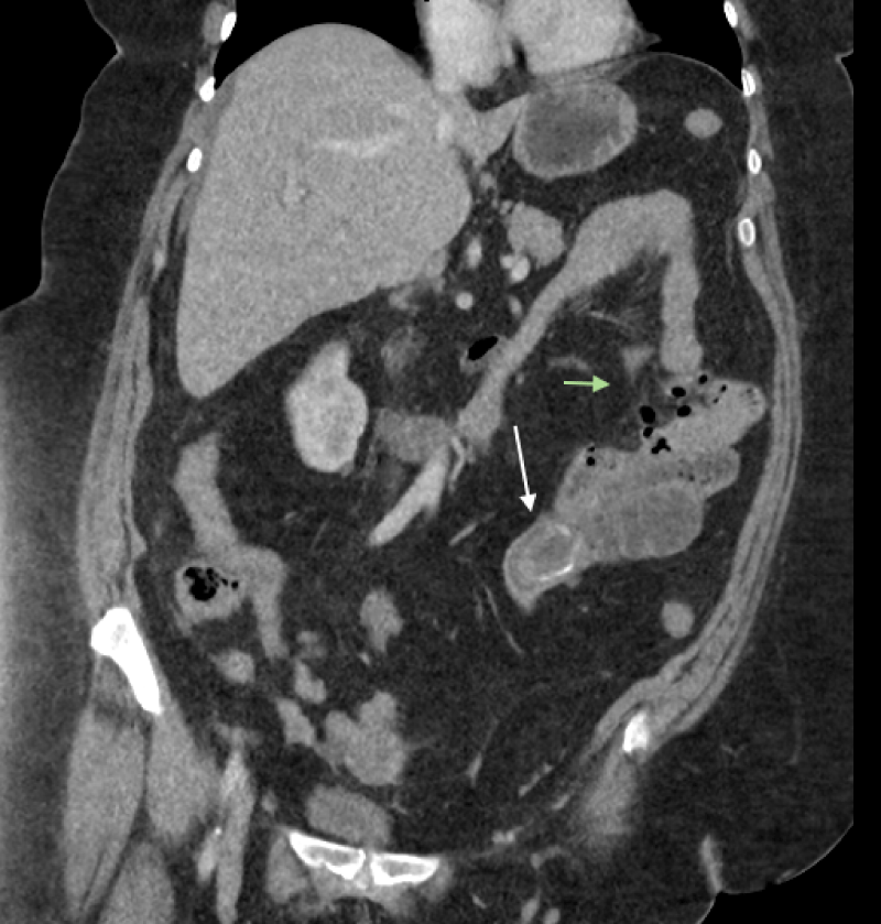
Figure 1:
Coronal computed tomography (CT) scan image demonstrating an ectopic gallstone (white arrow) in the proximal ileum and jejunal perforation (green arrow).
Read Full Article HTML DOI: 10.29328/journal.acr.1001050 Cite this Article Read Full Article PDF
More Images
Similar Articles
-
Trichomonas Vaginalis-A Clinical ImageAstrit M Gashi*. Trichomonas Vaginalis-A Clinical Image. . 2017 doi: 10.29328/journal.hjcr.1001001; 1: 001-002
-
Gastric Mucosal CalcinosisVedat Goral*,Irem Ozover,Ilknur Turkmen. Gastric Mucosal Calcinosis. . 2017 doi: 10.29328/journal.hjcr.1001002; 1: 003-005
-
Giant Lipoma Anterior Neck: A case reportManu CB*,Gowri Sankar M,Arun Alexander. Giant Lipoma Anterior Neck: A case report . . 2017 doi: 10.29328/journal.hjcr.1001003; 1: 006-008
-
Catamenial pneumothorax: Presentation of an uncommon PathologyRui Haddad*,Caterin Arévalo,David Nigri. Catamenial pneumothorax: Presentation of an uncommon Pathology . . 2017 doi: 10.29328/journal.hjcr.1001004; 1: 009-013
-
A rare case: Congenital Megalourethra in prune belly syndromeNuman Baydilli*,Ismail Selvi,Emre Can Akınsal. A rare case: Congenital Megalourethra in prune belly syndrome. . 2018 doi: 10.29328/journal.acr.1001005; 2: 001-003
-
Trauma to the neck: Manifestation of injuries outside the original zone of injury-A case reportStephen O Heard*,Alexander Christakis,Brian Tashjian,Anne M Gilroy3. Trauma to the neck: Manifestation of injuries outside the original zone of injury-A case report. . 2018 doi: 10.29328/journal.acr.1001006; 2: 004-009
-
Meige Trofoedema: A form of primary lymphedemaCarlos Al Sanchez Salguero*. Meige Trofoedema: A form of primary lymphedema. . 2018 doi: 10.29328/journal.acr.1001007; 2: 010-014
-
A rare case of Diabetic Foot in male of middle age has been shown Diabetic footSujit K Bhattacharya*. A rare case of Diabetic Foot in male of middle age has been shown Diabetic foot. . 2018 doi: 10.29328/journal.acr.1001008; 2: 015-015
-
Brooke-Spiegler Syndrome: A rare cause of skin appendageal tumorN Suganthan*,S Pirasath,DD Dikowita. Brooke-Spiegler Syndrome: A rare cause of skin appendageal tumor. . 2018 doi: 10.29328/journal.acr.1001009; 2: 016-018
-
McArdle’s Disease (Glycogen Storage Disease type V): A Clinical CaseCameselle-Teijeiro JF*,Calheiros-Cruz T,Caamaño-Vara MP,Villar-Fernández B,Ruibal-Azevedo J,Cameselle-Cortizo L,Cameselle-Arias M,Charro Gamallo ME,Turienzo-Pacho F,Yera Acosta A. McArdle’s Disease (Glycogen Storage Disease type V): A Clinical Case. . 2018 doi: 10.29328/journal.acr.1001010; 2: 019-023
Recently Viewed
-
Evaluation of In vitro and Ex vivo Models for Studying the Effectiveness of Vaginal Drug Systems in Controlling Microbe Infections: A Systematic ReviewMohammad Hossein Karami*, Majid Abdouss*, Mandana Karami. Evaluation of In vitro and Ex vivo Models for Studying the Effectiveness of Vaginal Drug Systems in Controlling Microbe Infections: A Systematic Review. Clin J Obstet Gynecol. 2023: doi: 10.29328/journal.cjog.1001151; 6: 201-215
-
A General Evaluation of the Cellular Role in Drug Release: A Clinical Review StudyMohammad Hossein Karami* and Majid Abdouss*. A General Evaluation of the Cellular Role in Drug Release: A Clinical Review Study. Clin J Obstet Gynecol. 2024: doi: 10.29328/journal.cjog.1001162; 7: 042-050
-
Strengthening Healthcare Delivery in the Democratic Republic of Congo through Adequate Nursing WorkforceMin Kyung Shin, Tshibambe N Tshimbombu*. Strengthening Healthcare Delivery in the Democratic Republic of Congo through Adequate Nursing Workforce. Clin J Nurs Care Pract. 2024: doi: 10.29328/journal.cjncp.1001051; 8: 007-010
-
Factors Influencing Knowledge on the Completion of Treatment among Tuberculosis Patients under Directly Observed Treatment Strategy (DOTS) in a Selected Health Facility, the BahamasEsther S Daniel*, Latasha Collie, Alice Neymour, Nicole KA Martin-Chen, Kevin Moss, Kathy-Ann Lootawan, Virginia M Victor. Factors Influencing Knowledge on the Completion of Treatment among Tuberculosis Patients under Directly Observed Treatment Strategy (DOTS) in a Selected Health Facility, the Bahamas. Clin J Nurs Care Pract. 2024: doi: 10.29328/journal.cjncp.1001053; 8: 022-030
-
Proportion of Risk of Stroke among Male Patients Attending Outpatient Department of a Tertiary Care Hospital, Ernakulam District, KeralaBetcy George*, Susan Mathai, Ansha Mol Shaji, Christy Elsa Abraham, Jisna Johnson. Proportion of Risk of Stroke among Male Patients Attending Outpatient Department of a Tertiary Care Hospital, Ernakulam District, Kerala. Clin J Nurs Care Pract. 2024: doi: 10.29328/journal.cjncp.1001054; 8: 031-035
Most Viewed
-
Evaluation of Biostimulants Based on Recovered Protein Hydrolysates from Animal By-products as Plant Growth EnhancersH Pérez-Aguilar*, M Lacruz-Asaro, F Arán-Ais. Evaluation of Biostimulants Based on Recovered Protein Hydrolysates from Animal By-products as Plant Growth Enhancers. J Plant Sci Phytopathol. 2023 doi: 10.29328/journal.jpsp.1001104; 7: 042-047
-
Feasibility study of magnetic sensing for detecting single-neuron action potentialsDenis Tonini,Kai Wu,Renata Saha,Jian-Ping Wang*. Feasibility study of magnetic sensing for detecting single-neuron action potentials. Ann Biomed Sci Eng. 2022 doi: 10.29328/journal.abse.1001018; 6: 019-029
-
Physical activity can change the physiological and psychological circumstances during COVID-19 pandemic: A narrative reviewKhashayar Maroufi*. Physical activity can change the physiological and psychological circumstances during COVID-19 pandemic: A narrative review. J Sports Med Ther. 2021 doi: 10.29328/journal.jsmt.1001051; 6: 001-007
-
Pediatric Dysgerminoma: Unveiling a Rare Ovarian TumorFaten Limaiem*, Khalil Saffar, Ahmed Halouani. Pediatric Dysgerminoma: Unveiling a Rare Ovarian Tumor. Arch Case Rep. 2024 doi: 10.29328/journal.acr.1001087; 8: 010-013
-
Prospective Coronavirus Liver Effects: Available KnowledgeAvishek Mandal*. Prospective Coronavirus Liver Effects: Available Knowledge. Ann Clin Gastroenterol Hepatol. 2023 doi: 10.29328/journal.acgh.1001039; 7: 001-010

HSPI: We're glad you're here. Please click "create a new Query" if you are a new visitor to our website and need further information from us.
If you are already a member of our network and need to keep track of any developments regarding a question you have already submitted, click "take me to my Query."


