Figure 1
Oncocytic papillary cystadenoma of right laryngeal ventricle
Tomasz Ścięgosz*, Renata Kwiatek, Izabela El-Hassanieh and Piotr Ziółkowski
Published: 30 April, 2021 | Volume 5 - Issue 1 | Pages: 012-013
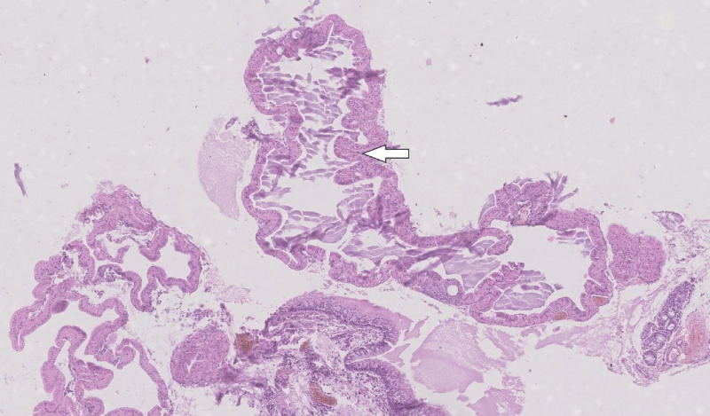
Figure 1:
The lesion formed multiple well-defined cysts with amorphous contents and short intraluminal papillary projections (white arrow). Unchanged laryngeal epithelium and seromucinous gland are also pictured. HE staining, 50x.
Read Full Article HTML DOI: 10.29328/journal.acr.1001048 Cite this Article Read Full Article PDF
More Images
Similar Articles
-
Clinical, histopathological and surgical evaluations of persistent oropharyngeal membrane case in a calfVehbi Gunes*,Gultekin Atalan,Latife Cakir Bayram,Kemal Varol,Hanifi Erol,Ihsan Keles,Ali C Onmaz. Clinical, histopathological and surgical evaluations of persistent oropharyngeal membrane case in a calf. . 2019 doi: 10.29328/journal.acr.1001016; 3: 021-025
-
Epiphora as a sign of unexpected underlying squamous cell carcinoma within sinonasal inverted papillomaFilippo Confalonieri*,Alessandra Di Maria,Raffaele Piscopo,Laura Balia,Luca Malvezzi. Epiphora as a sign of unexpected underlying squamous cell carcinoma within sinonasal inverted papilloma. . 2020 doi: 10.29328/journal.acr.1001038; 4: 038-040
-
Oncocytic papillary cystadenoma of right laryngeal ventricleTomasz Ścięgosz*,Renata Kwiatek,Izabela El-Hassanieh,Piotr Ziółkowski. Oncocytic papillary cystadenoma of right laryngeal ventricle. . 2021 doi: 10.29328/journal.acr.1001048; 5: 012-013
-
Endoscopic Endonasal total Removal of a Suprasellar, Preinfundibular Retro Chiasmatic Craniopharyngioma: A Surgical Case ReportAlessandra Alfieri, Armando Rapanà, Ferdinando Caranci. Endoscopic Endonasal total Removal of a Suprasellar, Preinfundibular Retro Chiasmatic Craniopharyngioma: A Surgical Case Report. . 2024 doi: 10.29328/journal.acr.1001090; 8: 036-038
Recently Viewed
-
Impact of Balanced Lifestyles on Childhood Development: A Study at CrècheP Vasundhara,P Nagaraju*. Impact of Balanced Lifestyles on Childhood Development: A Study at Crèche. J Addict Ther Res. 2024: doi: 10.29328/journal.jatr.1001028; 8: 001-008
-
Relationship between the Level of Spirituality and Blood Pressure Control among Adult Hypertensive Patients in a Southwestern Community in NigeriaAdetunji OMONIJO*, Paul OLOWOYO, Azeez Oyemomi IBRAHIM, Segun Matthew AGBOOLA, Oluwaserimi Adewumi AJETUNMOBI, Temitope Moronkeji OLANREWAJU, Adejumoke Oluwatosin OMONIJO. Relationship between the Level of Spirituality and Blood Pressure Control among Adult Hypertensive Patients in a Southwestern Community in Nigeria. Ann Clin Hypertens. 2023: doi: 10.29328/journal.ach.1001034; 7: 004-012
-
Progress in the development of Lipoplex and Polyplex modified with Anionic Polymer for efficient Gene DeliveryYoshiyuki Hattori*. Progress in the development of Lipoplex and Polyplex modified with Anionic Polymer for efficient Gene Delivery. J Genet Med Gene Ther. 2017: doi: 10.29328/journal.jgmgt.1001002; 1: 003-018
-
The advances and challenges of Gene Therapy for Duchenne Muscular DystrophyJacques P Tremblay*,Jean-Paul Iyombe-Engembe. The advances and challenges of Gene Therapy for Duchenne Muscular Dystrophy. J Genet Med Gene Ther. 2017: doi: 10.29328/journal.jgmgt.1001003; 1: 019-036
-
Management and Therapeutic Strategies for Spinal Muscular AtrophySheena P Kochumon, Cherupally Krishnan Krishnan Nair*. Management and Therapeutic Strategies for Spinal Muscular Atrophy. J Genet Med Gene Ther. 2024: doi: 10.29328/journal.jgmgt.1001009; 7: 001-007
Most Viewed
-
Evaluation of Biostimulants Based on Recovered Protein Hydrolysates from Animal By-products as Plant Growth EnhancersH Pérez-Aguilar*, M Lacruz-Asaro, F Arán-Ais. Evaluation of Biostimulants Based on Recovered Protein Hydrolysates from Animal By-products as Plant Growth Enhancers. J Plant Sci Phytopathol. 2023 doi: 10.29328/journal.jpsp.1001104; 7: 042-047
-
Feasibility study of magnetic sensing for detecting single-neuron action potentialsDenis Tonini,Kai Wu,Renata Saha,Jian-Ping Wang*. Feasibility study of magnetic sensing for detecting single-neuron action potentials. Ann Biomed Sci Eng. 2022 doi: 10.29328/journal.abse.1001018; 6: 019-029
-
Physical activity can change the physiological and psychological circumstances during COVID-19 pandemic: A narrative reviewKhashayar Maroufi*. Physical activity can change the physiological and psychological circumstances during COVID-19 pandemic: A narrative review. J Sports Med Ther. 2021 doi: 10.29328/journal.jsmt.1001051; 6: 001-007
-
Evaluation of In vitro and Ex vivo Models for Studying the Effectiveness of Vaginal Drug Systems in Controlling Microbe Infections: A Systematic ReviewMohammad Hossein Karami*, Majid Abdouss*, Mandana Karami. Evaluation of In vitro and Ex vivo Models for Studying the Effectiveness of Vaginal Drug Systems in Controlling Microbe Infections: A Systematic Review. Clin J Obstet Gynecol. 2023 doi: 10.29328/journal.cjog.1001151; 6: 201-215
-
Pediatric Dysgerminoma: Unveiling a Rare Ovarian TumorFaten Limaiem*, Khalil Saffar, Ahmed Halouani. Pediatric Dysgerminoma: Unveiling a Rare Ovarian Tumor. Arch Case Rep. 2024 doi: 10.29328/journal.acr.1001087; 8: 010-013

HSPI: We're glad you're here. Please click "create a new Query" if you are a new visitor to our website and need further information from us.
If you are already a member of our network and need to keep track of any developments regarding a question you have already submitted, click "take me to my Query."


