Figure 1
CT perfusion-guided endovascular treatment of symptomatic cerebral vasospasm in a patient with perimesencephalic non-aneurysmal subarachnoid hemorrhage
Andrea Saletti*, Andrea Bernardoni, Luca Borgatti1, Giuseppe Carità, Marco Farneti, Onofrio Marcello and Enrico Fainardi
Published: 31 March, 2020 | Volume 4 - Issue 1 | Pages: 020-021
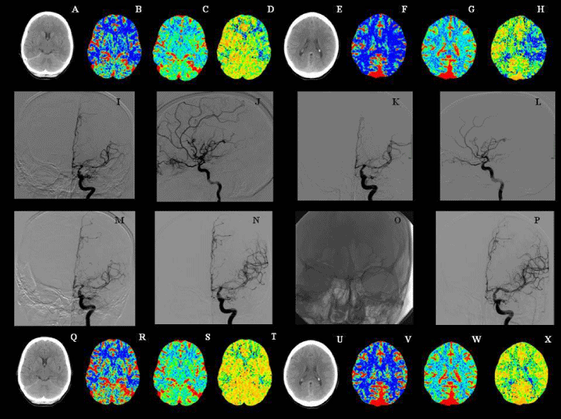
Figure 1:
Figure A-H: Standard CT scan and cerebral blood flow (CBF), cerebral blood volume (CBV) and mean transit time (MTT) CTP maps obtained 11 days after admission at the level of basal ganglia (A-D) and lateral ventricles (E-H) and showing reduced CBF, increased MTT and normal CBV in the left middle cerebral artery (MCA) territory, in absence of focal areas of hypoattenuation. Figure I-L: the following cerebral digital subtraction angiography (DSA) demonstrating a severe vasospasm in sovraclinoid segment of both internal carotid artery and in the M1 segment of left MCA (I-J) and its partial remission observed after the intra-arterial injection of nimodipine (K-L). Figure M-P: cerebral DSA performed 12 days after entry and illustrating the recrudescence of vasospasm (M), its initial improvement after nimodipine intra-arterial administration (N), balloon positioning in the left MCA (O) and the final resolution of arterial narrowing (P). Figure Q-X: Standard CT scan and CBF, CBV and MTT CTP maps acquired 13 days after admission at the level of basal ganglia (Q-T) and lateral ventricles (U-X) and documenting the complete restoring of cerebral perfusion in the left MCA territory.
Read Full Article HTML DOI: 10.29328/journal.acr.1001033 Cite this Article Read Full Article PDF
More Images
Similar Articles
-
Trichomonas Vaginalis-A Clinical ImageAstrit M Gashi*. Trichomonas Vaginalis-A Clinical Image. . 2017 doi: 10.29328/journal.hjcr.1001001; 1: 001-002
-
Gastric Mucosal CalcinosisVedat Goral*,Irem Ozover,Ilknur Turkmen. Gastric Mucosal Calcinosis. . 2017 doi: 10.29328/journal.hjcr.1001002; 1: 003-005
-
Giant Lipoma Anterior Neck: A case reportManu CB*,Gowri Sankar M,Arun Alexander. Giant Lipoma Anterior Neck: A case report . . 2017 doi: 10.29328/journal.hjcr.1001003; 1: 006-008
-
Catamenial pneumothorax: Presentation of an uncommon PathologyRui Haddad*,Caterin Arévalo,David Nigri. Catamenial pneumothorax: Presentation of an uncommon Pathology . . 2017 doi: 10.29328/journal.hjcr.1001004; 1: 009-013
-
A rare case: Congenital Megalourethra in prune belly syndromeNuman Baydilli*,Ismail Selvi,Emre Can Akınsal. A rare case: Congenital Megalourethra in prune belly syndrome. . 2018 doi: 10.29328/journal.acr.1001005; 2: 001-003
-
Trauma to the neck: Manifestation of injuries outside the original zone of injury-A case reportStephen O Heard*,Alexander Christakis,Brian Tashjian,Anne M Gilroy3. Trauma to the neck: Manifestation of injuries outside the original zone of injury-A case report. . 2018 doi: 10.29328/journal.acr.1001006; 2: 004-009
-
Meige Trofoedema: A form of primary lymphedemaCarlos Al Sanchez Salguero*. Meige Trofoedema: A form of primary lymphedema. . 2018 doi: 10.29328/journal.acr.1001007; 2: 010-014
-
A rare case of Diabetic Foot in male of middle age has been shown Diabetic footSujit K Bhattacharya*. A rare case of Diabetic Foot in male of middle age has been shown Diabetic foot. . 2018 doi: 10.29328/journal.acr.1001008; 2: 015-015
-
Brooke-Spiegler Syndrome: A rare cause of skin appendageal tumorN Suganthan*,S Pirasath,DD Dikowita. Brooke-Spiegler Syndrome: A rare cause of skin appendageal tumor. . 2018 doi: 10.29328/journal.acr.1001009; 2: 016-018
-
McArdle’s Disease (Glycogen Storage Disease type V): A Clinical CaseCameselle-Teijeiro JF*,Calheiros-Cruz T,Caamaño-Vara MP,Villar-Fernández B,Ruibal-Azevedo J,Cameselle-Cortizo L,Cameselle-Arias M,Charro Gamallo ME,Turienzo-Pacho F,Yera Acosta A. McArdle’s Disease (Glycogen Storage Disease type V): A Clinical Case. . 2018 doi: 10.29328/journal.acr.1001010; 2: 019-023
Recently Viewed
-
Beta-1 Receptor (β1) in the Heart Specific Indicate to StereoselectivityRezk Rezk Ayyad*, Ahmed Mohamed Mansour, Ahmed Mohamed Nejm, Yasser Abdel Allem Hassan, Norhan Hassan Gabr, Ahmed Rezk Ayyad. Beta-1 Receptor (β1) in the Heart Specific Indicate to Stereoselectivity. Arch Pharm Pharma Sci. 2024: doi: 10.29328/journal.apps.1001060; 8: 082-088
-
Schizoaffective Disorder in an Individual with Mowat-Wilson Syndrome (MWS)Yadwinder Chuhan, Nimrit Bath, Muhammad Ayub*. Schizoaffective Disorder in an Individual with Mowat-Wilson Syndrome (MWS). Arch Psychiatr Ment Health. 2024: doi: 10.29328/journal.apmh.1001050; 8: 008-011
-
Mapping the Psychosocial: Introducing a Standardised System to Improve Psychosocial Understanding within Mental HealthMatthew Bretton Oakes*. Mapping the Psychosocial: Introducing a Standardised System to Improve Psychosocial Understanding within Mental Health. Arch Psychiatr Ment Health. 2024: doi: 10.29328/journal.apmh.1001051; 8: 012-019
-
Approaching Mental Health Through a Preventive Data Analysis PlatformGabriel F Pestana, Olga Valentim*. Approaching Mental Health Through a Preventive Data Analysis Platform. Arch Psychiatr Ment Health. 2024: doi: 10.29328/journal.apmh.1001052; 8: 020-027
-
Fifteen year Follow-up of Relapsed/ Refractory Patients with Hodgkin Lymphoma Treated with Autologous Hematopoietic Stem Cell TransplantationSabree Abredabo*, Harrah Chiang, Leonard Klein, Tulio Rodriguez, Jacob D Bitran. Fifteen year Follow-up of Relapsed/ Refractory Patients with Hodgkin Lymphoma Treated with Autologous Hematopoietic Stem Cell Transplantation. J Stem Cell Ther Transplant. 2024: doi: 10.29328/journal.jsctt.1001040; 8: 033-037
Most Viewed
-
Evaluation of Biostimulants Based on Recovered Protein Hydrolysates from Animal By-products as Plant Growth EnhancersH Pérez-Aguilar*, M Lacruz-Asaro, F Arán-Ais. Evaluation of Biostimulants Based on Recovered Protein Hydrolysates from Animal By-products as Plant Growth Enhancers. J Plant Sci Phytopathol. 2023 doi: 10.29328/journal.jpsp.1001104; 7: 042-047
-
Feasibility study of magnetic sensing for detecting single-neuron action potentialsDenis Tonini,Kai Wu,Renata Saha,Jian-Ping Wang*. Feasibility study of magnetic sensing for detecting single-neuron action potentials. Ann Biomed Sci Eng. 2022 doi: 10.29328/journal.abse.1001018; 6: 019-029
-
Physical activity can change the physiological and psychological circumstances during COVID-19 pandemic: A narrative reviewKhashayar Maroufi*. Physical activity can change the physiological and psychological circumstances during COVID-19 pandemic: A narrative review. J Sports Med Ther. 2021 doi: 10.29328/journal.jsmt.1001051; 6: 001-007
-
Evaluation of In vitro and Ex vivo Models for Studying the Effectiveness of Vaginal Drug Systems in Controlling Microbe Infections: A Systematic ReviewMohammad Hossein Karami*, Majid Abdouss*, Mandana Karami. Evaluation of In vitro and Ex vivo Models for Studying the Effectiveness of Vaginal Drug Systems in Controlling Microbe Infections: A Systematic Review. Clin J Obstet Gynecol. 2023 doi: 10.29328/journal.cjog.1001151; 6: 201-215
-
Pediatric Dysgerminoma: Unveiling a Rare Ovarian TumorFaten Limaiem*, Khalil Saffar, Ahmed Halouani. Pediatric Dysgerminoma: Unveiling a Rare Ovarian Tumor. Arch Case Rep. 2024 doi: 10.29328/journal.acr.1001087; 8: 010-013

HSPI: We're glad you're here. Please click "create a new Query" if you are a new visitor to our website and need further information from us.
If you are already a member of our network and need to keep track of any developments regarding a question you have already submitted, click "take me to my Query."


