
More Information
Submitted: March 18, 2021 | Approved: July 15, 2021 | Published: July 16, 2021
How to cite this article: Mehrotra S, Huang Y, Knittel R, Thirunavukkarasu P. Gallstone Ileus with associated perforated small bowel diverticulitis. Arch Case Rep. 2021; 5: 021-022.
DOI: 10.29328/journal.acr.1001050
Copyright License: © 2021 Mehrotra S, et al. This is an open access article distributed under the Creative Commons Attribution License, which permits unrestricted use, distribution, and reproduction in any medium, provided the original work is properly cited.
Keywords: Pterygoid implants; One piece implant; Edentulous patient; Rehabilitation

Gallstone Ileus with associated perforated small bowel diverticulitis
Shreeja Mehrotra*, Yang Huang, Ronan Knittel and Palan Thirunavukkarasu
Department of General Surgery, Sir Charles Gairdner Hospital, Perth, Western Australia, Australia
*Address for Correspondence: Dr. Shreeja Mehrotra. MB, ChB Department of General Surgery, Sir Charles Gairdner Hospital, Hospital Ave, Nedlands 6009 WA, Australia, Tel: +61 6457 3333; Email: [email protected]
Gallstone ileus is a rare complication of cholelithiasis and a type of mechanical obstruction involving impaction of a gallstone within the intestinal tract [1,2]. This entity occurs in 0.15% - 1.5% of cholelithiasis cases and < 0.1% of ileus cases overall [1]. Gallstone ileus is more common in the elderly and up to 80% - 90% of affected patients have medical comorbidities [2]. The ratio of occurrence in females to males is 3.5:1 [3].
The following report presents a case of gallstone ileus with associated perforated small bowel diverticulitis, demonstrating the importance of considering this condition as a differential diagnosis of an acute abdomen.
We present a case of a 72-year-old female patient who presented with sudden onset left iliac fossa pain associated with nausea and vomiting. This was on a background of a recent diagnosis of Mirizzi syndrome. Her comorbidities included a previous history of invasive ductal breast cancer one year previously, completed wide local excision and adjuvant radiotherapy, hypertension, rheumatoid arthritis and depression. On examination, she had mild abdominal distension with rebound tenderness in the left iliac fossa. Her bloods showed a mild leucocytosis, CRP of 32 and abnormal liver function tests with ALP of 263 and GGT of 1140.
The computed tomography (CT) scan demonstrated a cholecystenteric fistula with a shrunken gall bladder containing a small amount of air. Pneumobilia was evident involving the common bile duct. Features of small bowel obstruction were visible with a transition point at the left lower quadrant, at which point an oval lamellated structure indicative of an ectopic gallstone was seen (Figure 1, white arrow). Collectively, these findings were consistent with the Rigler triad for gallstone ileus (pneumobilia, small bowel obstruction and ectopic gallstone). Incidentally, CT analysis also revealed evidence of small bowel diverticulitis within a 5 cm segment proximal to the impacted gallstone (Figure 1, green arrow).
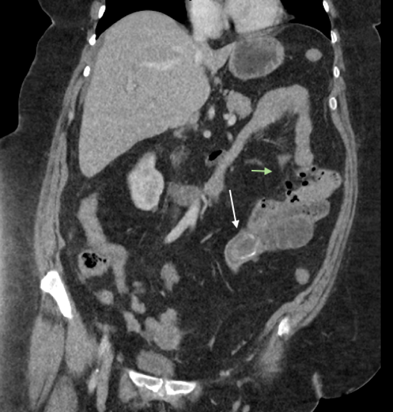
Figure 1: Coronal computed tomography (CT) scan image demonstrating an ectopic gallstone (white arrow) in the proximal ileum and jejunal perforation (green arrow).
The patient underwent an uncomplicated laparotomy, removal of gallstone and small bowel resection. Intra-operatively, a 6.7 cm gallstone (Figure 1) was found impacted in the proximal ileum. Two small jejunal diverticulae were noted, one of which had perforated (Figure 1) secondary to increased intraluminal pressure from gallstone ileus. The gallstone was mechanically migrated proximally to the site of the small bowel diverticulitis perforation and a 10 cm segment of small bowel was resected, with the gallstone retrieved via the enterotomy opening. Her post-operative course was uneventful and she was discharged day 6 post operatively. The patient was noted to have progressed well at post-operative clinic follow-up, as well as six-month telephone follow-up.
Gallstone ileus is often preceded by an episode of acute cholecystitis and the inflammation and pressure exerted from the offending gallstone leads to erosion through the gallbladder wall. This results in fistula formation between the gallbladder and the adjacent and adhered portion of the gastrointestinal tract, allowing passage of the gallstone [2].
The clinical signs and symptoms of gallstone ileus are usually non-specific, which can contribute to a delay in diagnosis [4]. Plain abdominal x-rays may show features of Rigler’s triad, however the sensitivity of plain abdominal radiographs alone in diagnosing gallstone ileus is poor (40% - 70%) [4]. Contrast-enhanced CT has been reported to have high sensitivity (93%), specificity (100%) and accuracy (99%) in diagnosing gallstone ileus [2].
In the presented case, the CT was crucial to diagnosis of gallstone ileus given the non-specific symptoms, therefore this case highlights the importance of its use.
Surgical management of gallstone ileus involves stone extraction to relieve intestinal obstruction and closure of the fistula. Another consideration is about the need for elective cholecystectomy [5]. The main controversy regarding surgical intervention exists around whether biliary surgery is performed at the same time as stone extraction (one-stage procedure), or whether this is carried out at a later stage (two-stage procedure), or not done at all [5]. Rarely, there may be spontaneous resolution of gallstone ileus with patients passing the stones via the rectum [3].
Gallstone ileus with proximal small bowel perforation is very rare [6]. In particular, there have been very few reports of gallstone ileus complicated by jejunal perforation [6,7]. Perforation can occur at the site of impaction or previous sites of obstruction due to pressure necrosis of the jejunal wall due to the gallstone. Alternatively, as highlighted in our case, perforation of a pre-existing jejunal diverticulum may occur [6,8]. The prevalence of jejunal diverticula is approximately 1% in the general population and it affects a similar age group to gallstone ileus, with its prevalence increasing in the elderly and peaking in the sixth and seventh decades [8]. Perforation of a jejunal diverticulum may occur in gallstone ileus secondary to increased intraluminal pressure from the obstructing stone [8].
The presented case highlights the importance of consideration of perforated small bowel diverticulitis as an associated finding with gallstone ileus.
While gallstone ileus is a rare complication of cholelithiasis, given the high prevalence of gallstones in the general population [1], this case highlights the importance of considering gallstone ileus as a differential diagnosis of an acute abdomen. Another key learning point from this report is to consider the possibility of associated perforated small bowel diverticulitis, as this is a rare potential complication of gallstone ileus.
- Inukai K. Gallstone ileus: a review. BMJ Open Gastroenterol. 2019; 6: e000344. PubMed:https://pubmed.ncbi.nlm.nih.gov/31875141/
- Nuno-Guzman CM, Marin-Contreras ME, Figueroa-Sanchez M, Corona JL. Gallstone ileus, clinical presentation, diagnostic and treatment approach. World J Gastrointest Surg. 2016; 8: 65-76. PubMed: https://pubmed.ncbi.nlm.nih.gov/26843914/
- Takahashi K, Kashimura H, Konno N, Nakagawa M, Kawahara Y, et al. Gallstone ileus with spontaneous evacuation: A case report. J Gen Fam Med. 2018; 19: 173-175. PubMed: https://pubmed.ncbi.nlm.nih.gov/30186731/
- Ravikumar R, Williams JG. The operative management of gallstone ileus. Ann R Coll Surg Engl. 2010; 92: 279-281. PubMed: https://pubmed.ncbi.nlm.nih.gov/20501012/
- Abou-Saif A, Al-Kawas FH. Complications of gallstone disease: Mirizzi syndrome, cholecystocholedochal fistula, and gallstone ileus. Am J Gastroenterol. 2002; 97: 249-254. PubMed: https://pubmed.ncbi.nlm.nih.gov/11866258/
- Lee CH, Yin WY, Chen JH. Gallstone ileus with jejunum perforation managed with laparoscopic-assisted surgery: rare case report and minimal invasive management. Int Surg. 2015; 100: 878-881. PubMed: https://pubmed.ncbi.nlm.nih.gov/26011209/
- Martin-Perez J, Bravo-Gutierrez A, Delgado-Plasencia L, Hernandez-Leon CN, Medina-Arana V. Jejunal diverticular perforation due to gallstone ileus. Rev Esp Enferm Dig. 2012; 104: 503-505. PubMed: https://pubmed.ncbi.nlm.nih.gov/23130865/
- Browning LE, Taylor JD, Clark SK, Karanjia ND. Jejunal perforation in gallstone ileus - a case series. J Med Case Rep. 2007; 1: 157. PubMed: https://pubmed.ncbi.nlm.nih.gov/18045463/