
More Information
Submitted: 14 April 2020 | Approved: 16 April 2020 | Published: 17 April 2020
How to cite this article: Wang Y, Zhou P, Tian X, Meng S, Sun X, et al. First cure case of 2019 novel coronavirus in Ningxia, China. Arch Case Rep. 2020; 4: 026-027.
DOI: 10.29328/journal.acr.1001035
Copyright License: © 2020 Wang Y, et al. This is an open access article distributed under the Creative Commons Attribution License, which permits unrestricted use, distribution, and reproduction in any medium, provided the original work is properly cited.

First cure case of 2019 novel coronavirus in Ningxia, China
Yifan Wang1, Pan Zhou2, Xingcang Tian1, Shuping Meng1, Xiao Sun1, Ting Li1 and Li Zhu1*
1Department of Radiology, General Hospital of Ningxia Medical University, China
2Department of Radiology, The Fourth People’s Hospital of Ningxia, China
*Address for Correspondence: Li Zhu, PhD, Professor, Department of Radiology, General Hospital of Ningxia Medical University, China, Tel: +86- 13995172918; Email: [email protected]
On January 19, 2020, a 28-year-old male presented to the hospital with a 2-day history of fever, occasional cough and headache. He disclosed that he worked in Wuhan [1], China (the center of novel coronavirus outbreak) and flew to Yinchuan on the day of admission.
The body temperature of the patient was elevated to 38.5°C (101.3°F, normal range 36.3-37.2 °C/97.34-98.96 °F). Coarse breath sounds were noted in both lungs on auscultation. Laboratory results showed white blood cell count was 4.37 × 10⁹/L (normal range, 3.5-9.5 × 10⁹/L), neutrophils were 52.8% (normal range, 40-75%) and lymphocytes were 35.9% (normal range, 20-50%); C-reactive protein was 0.55 mg/L (normal range, 0-6 mg/L). Prothrombin time was 11.4s (normal range, 10-14s), activated partial prothrombin time was 29.6s (normal range, 23-35s), Thrombin time was 20.3s (normal range, 13-25s) and D-dimer was 0.30 ug/mL (normal range, 0-1 ug/mL). Unenhanced chest computed tomography (CT) showed multiple patchy ground-glass opacities and consolidation in the right middle lobe and lower lobe of both lungs (Figure 1 A-C). The real-time reverse transcriptase polymerase chain reaction (RT-PCR) of the patient’s oropharyngeall swab specimens were positive for 2019 novel coronavirus (nCoV) nucleic acid.
Given epidemiological characteristics that the patient came from the infected area, clinical manifestations, CT findings and laboratory tests, the diagnosis of 2019-nCoV pneumonia was confirmed [2-3]. After receiving 14 days antiviral therapy with moxifloxacin (0.4 g administered intravenously per day), lopinavir/ritonavir (400/100 mg administered orally every 12 hours) and interferon inhalation (5 million IU every 12 hours), the patient clinically improved with obviously reduced pulmonary opacities found at repeat chest CT (Figure 1 D-F) and twice consecutive negative RT-PCR. The temperature returned to 36.5 °C (97.7 °F) from January 22, and the patient discharged on February 3rd. The patient was isolated at home for 14 days after checking out of hospital to monitor temperature every day.
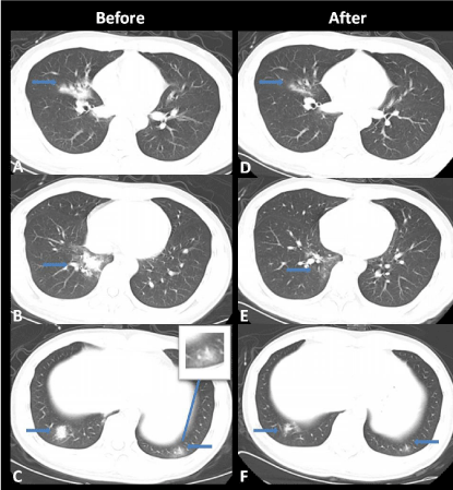
Figure 1: Thoracic non-contrast CT images in a 28-year-old male before and after treatment. (A-C), Image shows multiple patchy ground-glass opacities and consolidation in the right middle lobe and basal segments of both lower lobes. (D-F), Follow-up image on January 31 shows obviously reduced ground-glass opacities in the right middle lobe and lower lobes of both lungs. The pulmonary vascular thickening is typical CT sign of the 2019 novel coronavirus pneumonia.
Funding
This study was supported by Ningxia key research and development plan to support the special project (2020BEG03006) and Ningxia medical university science foundation (XM2018089).
Ethical approval
Patient’s data were anonymized.
- Li Q, Guan X, Wu P, Wang X, Zhou L, et al. Early transmission dynamics in Wuhan, China, of Novel coronavirus-infected pneumonia. N Engl J Med. 2020; 382: 1199-1207. PubMed: https://www.ncbi.nlm.nih.gov/pubmed/31995857
- Chen N, Zhou M, Dong X, Qu J, Gong F, et al. Epidemiological and clinical characteristics of 99 cases of 2019 novel coronavirus pneumonia in Wuhan, China: a descriptive study. Lancet. 2020; 395: 507-513. PubMed: https://www.ncbi.nlm.nih.gov/pubmed/32007143
- Huang C, Wang Y, Li X, Ren L, Zhao J, et al.Clinical features of patients infected with 2019 novel Coronavirus in Wuhan, China. Lancet.395: 497-506. PubMed: https://www.ncbi.nlm.nih.gov/pubmed/31986264