
More Information
Submitted: July 24, 2026 | Accepted: July 28, 2026 | Published: July 30, 2026
Citation: Torres-Gómez FJ, de Medina-González RS. Value of Systematic Histopathological Examination in Vaginal Reconstruction and Remodeling Specimens: Clinical, Diagnostic, and Quality Assurance. Arch Case Rep. 2026; 10(7): 74-76. Available from:
https://dx.doi.org/10.29328/journal.acr.1001192
DOI: 10.29328/journal.acr.1001192
Copyright license: © 2026 Torres-Gómez FJ, et al. This is an open access article distributed under the Creative Commons Attribution License, which permits unrestricted use, distribution, and reproduction in any medium, provided the original work is properly cited.

Value of Systematic Histopathological Examination in Vaginal Reconstruction and Remodeling Specimens: Clinical, Diagnostic, and Quality Assurance
Francisco Javier Torres-Gómez and Rosa Sánchez de Medina-González
Dr. Torres Laboratory of Pathology and Cytology (CITADIAG SL). Seville, Spain
*Corresponding author: Francisco Javier Torres-Gómez, Dr. Torres Laboratory of Pathology and Cytology (CITADIAG SL). Seville, Spain, Email: [email protected]
In daily surgical practice, clinicians and pathologists may encounter the temptation of assuming that tissue excised for purely anatomical, aesthetic, or functional reconstructive reasons lacks significant underlying pathology. While gross examination or clinical impression often suggests benignity, submitting surgical specimens for formal anatomical pathology evaluation represents an essential component of clinical thoroughness, objective verification, and risk management.
This discussion is illustrated by the routine histopathological examination of a surgical specimen from acolpoplasty and vaginal repair performed on a 70-year-old postmenopausal woman presenting with symptomatic stage III pelvic organ prolapse (POP; cystocele and rectocele). Preoperative cervical/vaginal cytology showed no intraepithelial lesions or malignancy. Following informed patientconsent, tissue was excised and submitted in 10% neutral buffered formalin. Gross evaluation showed atissue fragment measuring 2.2 × 2.0 × 1.3 cm in maximum dimensions, presenting a grayish-white outersurface and a soft-elastic consistency on sectioning.
Specimens were routinely processed, embedded in paraffin, sectioned at 4 µm, and stained with Hematoxylin and Eosin (H&E). Microscopic examination revealed a physiological tissue architecture, fully congruent with the patient postmenopausal hormonal status (Figures 1 to 5). A stratified squamous epithelium exhibiting marked senile atrophic changes was observed, characterized by epithelial thinning,loss of intermediate and superficial cell maturation, and focal areas of reactive superficial hyperkeratosis/orthokeratosis (Figures 2 and 4). The underlying subepithelial stroma displayed marked,paucicellular, hyalinized collagenous fibrosis with thin-walled blood vessels. In the deeper strata, mature adipose tissue intermingled with the surrounding fibrous tissue. There was a strict absence of cellularatypia, atypical mitoses, dysplastic epithelial proliferation (squamous intraepithelial lesions [SIL] /vaginal intraepithelial neoplasia [VAIN]), or specific inflammatory infiltrates (Figures 1,3,and 5).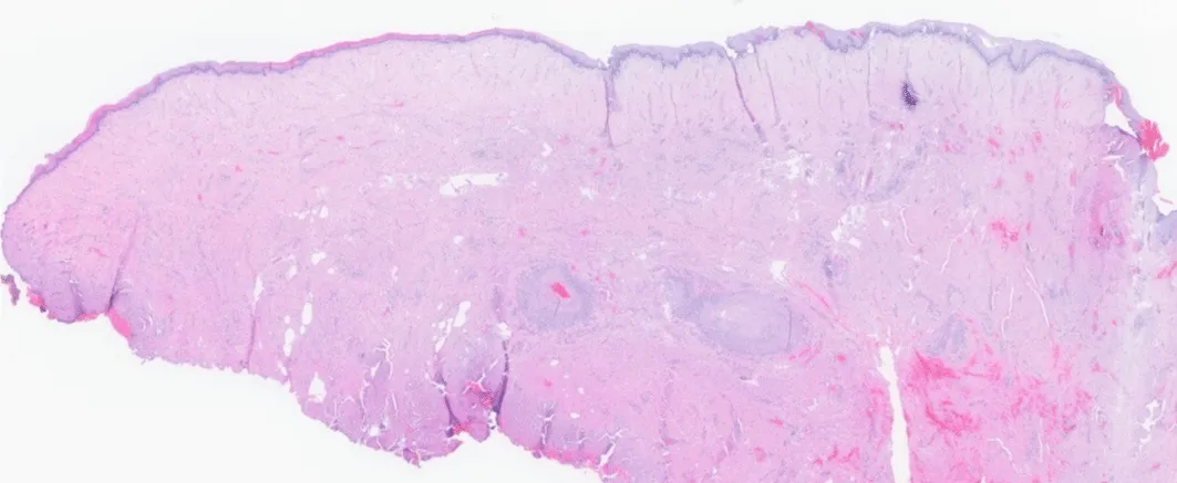
Figure 1: Histological overview of the vaginal repair specimen (H & amp; E, 2x objective magnification). Low-power view showing a tissue fragment lined by an atrophic stratified squamous epithelium. The underlying stroma exhibits marked fibrosis, dermal/submucosal hyalinization, and lobules of mature adipose tissue in the deep planes, without evidence of tumor proliferation or significant inflammatory infiltrates.
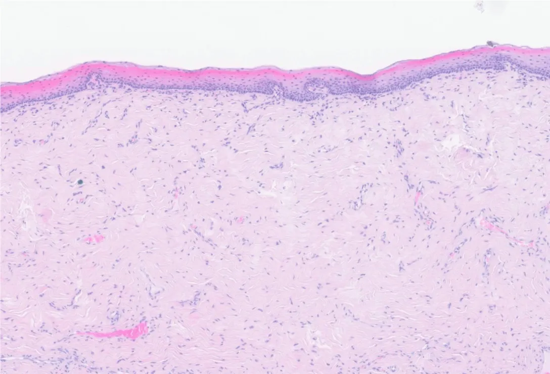
Figure 2: Atrophic squamous epithelium with focal keratinization (H&E, 10x objective magnification). Microscopic detail of the mucosal lining. Note the marked loss of thickness and maturation of the squamous epithelium (senile atrophic change) along with a thin, reactive, superficial hyperkeratotic/orthokeratotic layer. The underlying papillary and reticular stroma is dense and collagenized.
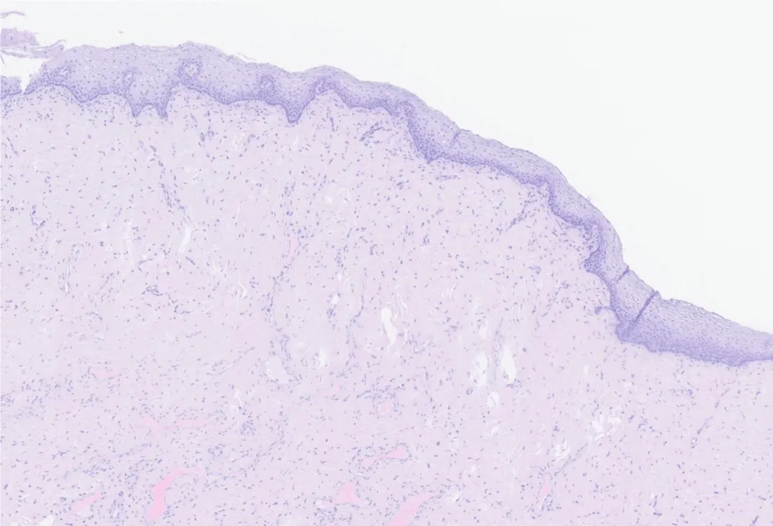
Figure 3: Mucocutaneous transition and stromal fibrosis (H&E, 10x objective magnification). Histological section of the mucosal margin displaying a thin, flattened, non-keratinized stratified squamous epithelium with blunting of the rete ridges. The lamina propria features a dense, paucicellular, well-vascularized fibrous stroma without nuclear atypia.
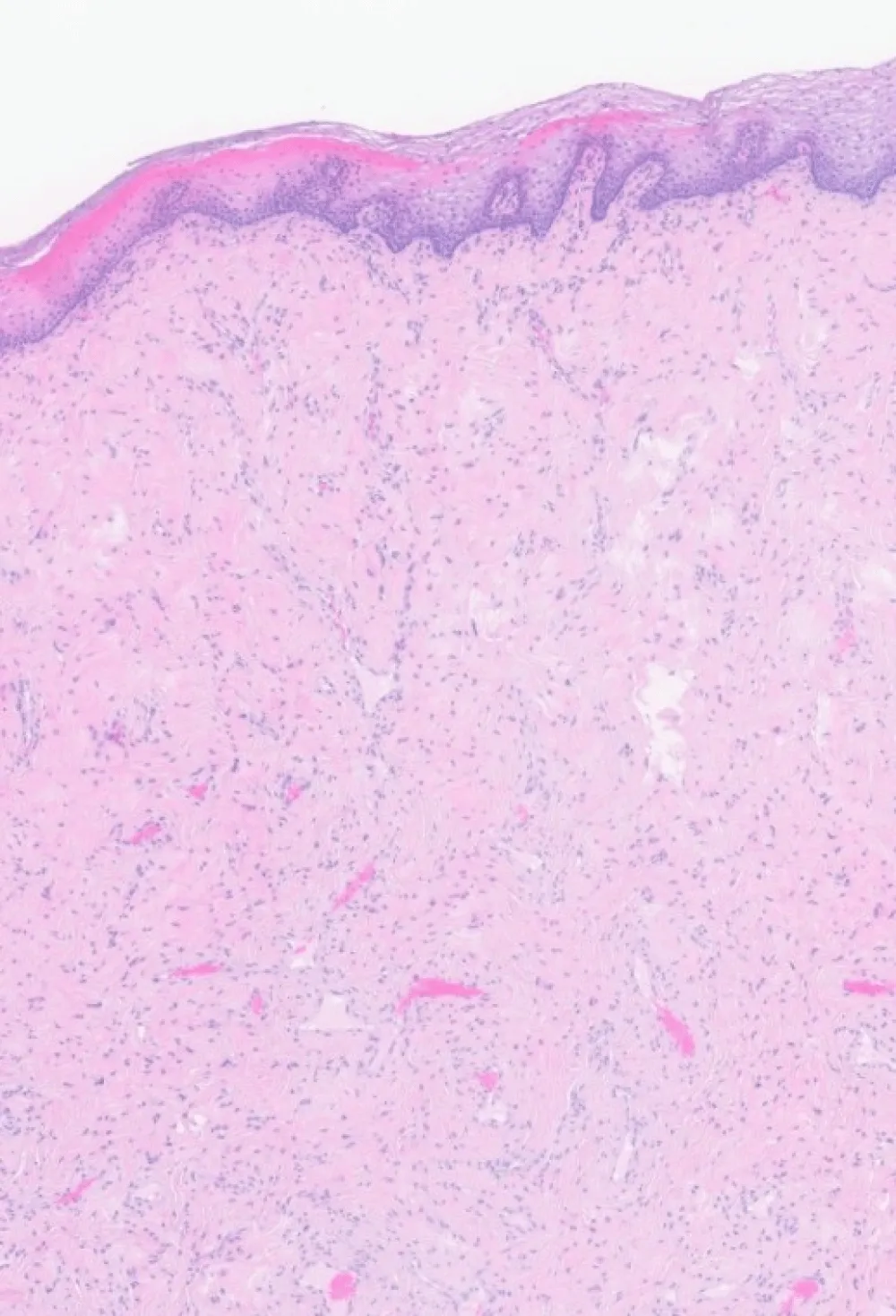
Figure 4: Detail of atrophic architecture and subepithelial stroma (H&E, 10x objective magnification). Preserved cellular polarity is observed within the atrophic squamous epithelium, topped by compact laminar orthokeratosis. Strict absence of dysplasia (VAIN/SIL), nuclear pleomorphism, or basal/suprabasal mitotic figures.
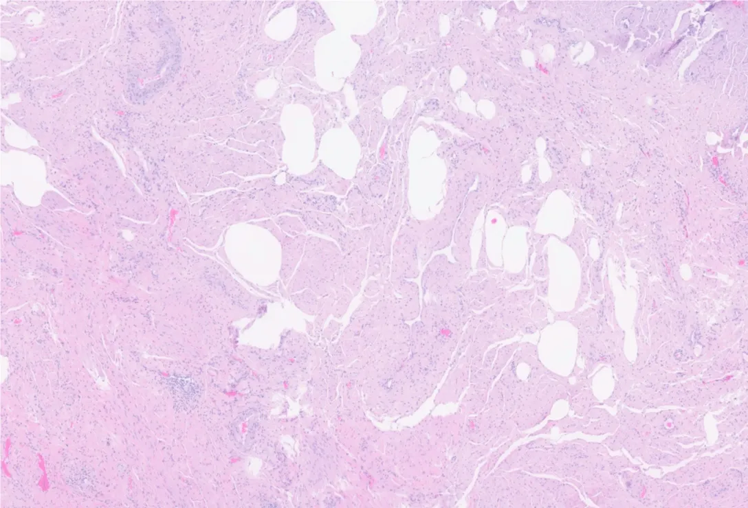
Figure 5: Deep stroma and fibroadipose component (H&E, 10x objective magnification). Section of the deep tissue body showing bands of dense, hyalinized connective tissue alternating with mature, non-atypical adipocytes and normally configured small-to-medium-caliber blood vessels. These findings areconsistent with age-related involutive/trophic remodeling.
In summary, the findings yielded the histopathological confirmation of benign stromal remodeling and involutive epithelial atrophy. Despite the absolute benignity of this case, a fundamental clinical question arises: to what extent is the systematic histopathological analysis of specimens obtained during reconstructive pelvic floor procedures (e.g., colpoplasties and POP repairs) or aesthetic gynecological surgeries (e.g., labioplasties) justified?
In recent decades, the debate regarding the cost-effectiveness of routine histological examination inbenign gynecological surgery has received attention in the medical literature. Several authors argue that the rate of unexpected malignant findings in such specimens is low, questioning the financial expenditureand clinical workload involved [1,2]. However, scientific literature documents clinically silent, unexpected histological findings in vaginal remodeling and prolapse resections. These range from incidental high-grade vaginal intraepithelial neoplasia (VAIN 3) and early-stage squamous cellcarcinomas [3], to insidious mesenchymal entities such as angiomyofibroblastoma, reactive fibroepithelialstromal tumors, or sarcoma botryoides /rhabdomyosarcoma presenting at atypical ages [4,5], as well asunrecognized deep infiltrative endometriosis, granulomatous infections (e.g., tuberculosis, deep fungal infections), or stromal amyloidosis deposits [6].
Comparing our specimen with these literature-reported findings illustrates how the histological spectrum of the vaginal wall spans from benign senile fibrous atrophy—such as the one presented here—to subclinical diagnostic traps. Without systematic microscopic examination, such conditions would pass unnoticed, potentially compromising patient management and long-term outcomes.
From a clinical and methodological perspective, foregoing microscopic evaluation converts visual Clinical judgment into an unverified assumption. Histopathological evaluation provides four distinct Clinical benefits: (1) diagnostic confirmation by excluding occult dysplasia or malignancy; (2) quality assurance of surgical care through objective tissue verification; (3) medico-legal protection documenting tissue benignity; and (4) patient safety mitigation against subclinical neoplastic or inflammatory processes.
Furthermore, distinguishing between functional reconstructive operations (colpoplasties/POP repair) and aesthetic procedures (labioplasties) is essential when assessing risk profiles across different patient demographics.
In conclusion, the case of our 70-year-old patient offers confirmation of tissue benignity. Nevertheless, it reinforces the pathological principle that patient safety should rely on objective microscopic evidence rather than visual impression alone. We advocate for maintaining the systematic histopathological evaluation of all vaginal plastic and reconstructive surgical specimens as an indispensable pillar ofdiagnostic accuracy and clinical safety in gynecology.
Ethics and patient consent
Written informed consent was obtained from the patient for publication of this case and accompanying images. In accordance with institutional guidelines, single retrospective case reviews of routine pathological specimens are exempt from formal institutional review board approval.
- Radaelli F, et al. Is routine pathological examination of vaginal wall tissue excised during pelvic organ prolapse surgery necessary? Int Urogynecol J. 2019;30(8):1321-1326.
- Dellas A, et al. Histopathological findings in vaginal wall resections during vaginal repair operations: a routine procedure under debate. Arch Gynecol Obstet. 2014;290(3):485-489.
- Boreham MK, et al. Incidental histological findings during pelvic organ prolapse surgery. Am J Obstet Gynecol. 2002;187(4):911-915.
- McCluggage WG. Mesenchymal tumors of the female genital tract: an update on selected benign andmalignant entities. Int J Gynecol Pathol. 2012;31(1):21-35.
- Loverro G, et al. Unexpected histological findings after vaginal prolapse repair: clinical implicationsand management. Eur J Obstet Gynecol Reprod Biol. 2018;221:112-116.
- Teresi M, et al. Unsuspected pathology in vaginal tissue excised during pelvic reconstructive surgery: asystematic review. Gynecol Surg. 2021;18(1):14.