
More Information
Submitted: July 08, 2026 | Accepted: July 13, 2026 | Published: July 15, 2026
Citation: Noorsher Z, Immers L, Torres Y, Ji SSG, Tagore KS. Bullous Pemphigoid Beyond the Skin: A Rare Cause of Oesophageal Haemorrhage. Arch Case Rep. 2026; 10(7): 71-73. Available from:
https://dx.doi.org/10.29328/journal.acr.1001191
DOI: 10.29328/journal.acr.1001191
Copyright license: © 2026 Noorsher Z, et al. This is an open access article distributed under the Creative Commons Attribution License, which permits unrestricted use, distribution, and reproduction in any medium, provided the original work is properly cited.
Keywords: Bullous pemphigoid, oesophageal haemorrhage, upper gastrointestinal bleeding, endoscopic Nikolsky sign

Bullous Pemphigoid Beyond the Skin: A Rare Cause of Oesophageal Haemorrhage
Zainab Noorsher1*, Lindsey Immers2, Yahanna Torres2, Samuel Shao Guang Ji3 and Kuldeep Singh Tagore3
1UC Davis School of Medicine, Sacramento, CA, USA
2Department of Internal Medicine, UC Davis Health, Sacramento, CA, USA
3Department of Gastroenterology and Hepatology, UC Davis Health, Sacramento, CA, USA
*Corresponding author: Zainab Noorsher, Medical Student, UC Davis School of Medicine, 4610 X Street, Sacramento, CA 95817, USA, Email: [email protected]
Background: Bullous pemphigoid (BP) is a chronic autoimmune bullous disease that predominantly affects elderly individuals with multiple comorbidities. Oesophageal involvement is uncommon, accounting for approximately 4% of cases. Oesophageal BP typically presents as chest pain, dysphagia, odynophagia or can remain entirely asymptomatic.
Case presentation: We report a 72-year-old female with a history of heart failure, atrial fibrillation on apixaban, and recently diagnosed BP on prednisone. She presented with acute hematemesis while sleeping and recurrent overnight blood-tinged expectoration. She had consumed hard nuts the previous night. She denied prior gastrointestinal bleeding or other risk factors.
Physical examination of the mouth revealed healing cutaneous BP lesions and dark red macules on the hard palate. Urgent esophagogastroduodenoscopy (EGD) revealed fragile, bullous oesophageal lesions. A tissue biopsy attempt was aborted due to immediate, excessive bleeding. Routine gastric biopsies revealed mild gastritis without malignancy or helicobacter pylori. No alternative bleeding sources were identified.
Throughout admission, the patient had no further haemorrhage or melena. Haemoglobin gradually stabilised at 11 g/dL without requiring transfusions. The hematemesis was attributed to mucosal injury of oesophageal BP lesions precipitated by mechanical trauma from hard food. Apixaban was safely resumed, and she was discharged on a high-dose proton pump inhibitor, a soft food diet, and her home maintenance BP therapy.
Conclusion: This case highlights the importance of suspecting mucosal BP in patients with upper gastrointestinal bleeding. Recognising this involvement allows for a modified procedural approach, ensuring cautious manipulation to mitigate the risk of severe, procedure-induced mucosal trauma or haemorrhage.
72-year-old female with a history of heart failure with reduced ejection fraction (on Bumex), atrial fibrillation (on apixaban), and bullous pemphigoid diagnosed six weeks prior and managed with prednisone and Dupilumab under dermatologic follow-up. The evening before presentation, she consumed pistachios and subsequently developed an episode of hematemesis during sleep while using her CPAP machine. She then experienced recurrent hematemesis and blood-tinged expectoration over the following five hours before presenting to the emergency department. She had no prior history of gastrointestinal bleeding and no known risk factors for upper GI bleed (UGIB) such as cirrhosis, GERD, or NSAID use.
On arrival, she was hypertensive and tachycardic. Initial haemoglobin was 13.7 g/dL. Physical examination was notable for diffuse pink to hyperpigmented patches on the trunk and extremities consistent with healing bullous pemphigoid lesions, without active tense bullae. Plantar skin sloughing and one to two pinpoint dark red macules on the hard palate of the mouth were also present.
Given concern for UGIB, upper endoscopy was performed, which revealed a bullous lesion within the oesophagus with marked mucosal fragility (Figure 1A). A single oesophageal lesion biopsy was attempted, which resulted in excessive bleeding and was treated with hemostatic powder (Figure 1B).
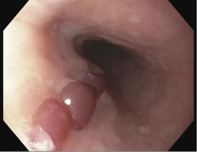
Figure 1A: Upper endoscopy findings demonstrating oesophageal bullous pemphigoid.
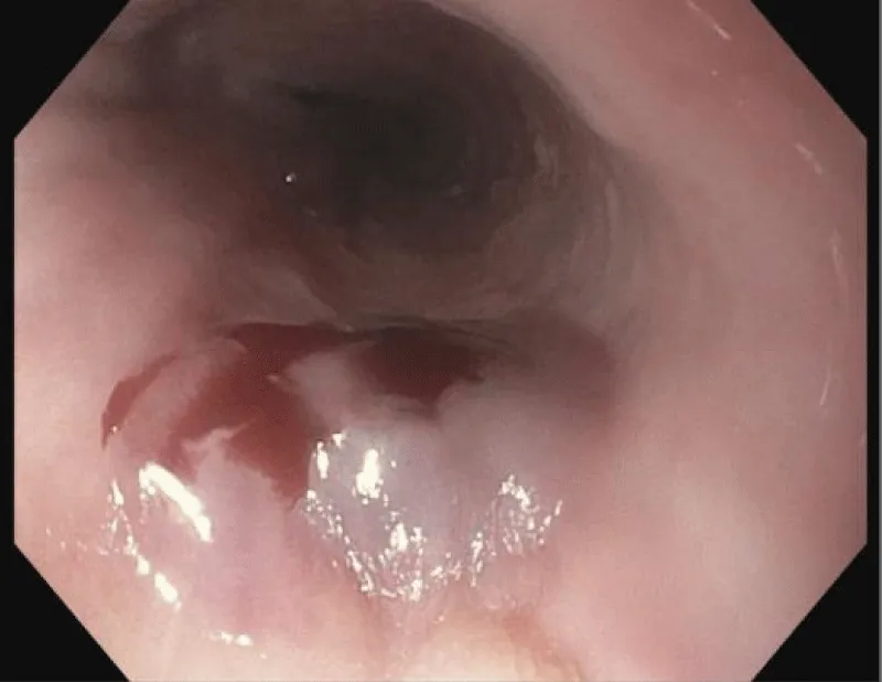
Figure 1B: Endoscopic view of the oesophagus demonstrating acute, biopsy-induced bleeding managed with immediate application of hemostatic powder.
Routine gastric biopsies revealed mild chronic inactive and chemical gastritis, but were negative for Helicobacter pylori, intestinal metaplasia, or dysplasia. No alternative source of UGIB was identified.
The patient had no further episodes of bleeding or melena, and Apixaban was resumed as she remained hemodynamically stable. Haemoglobin gradually decreased to approximately 11 g/dL without requiring transfusion. Given the absence of recurrent bleeding, no further interventions were made. Given the EGD findings and lack of alternative etiologies, the patient’s hematemesis was attributed to mucosal injury of oesophageal bullous pemphigoid lesions, possibly precipitated by mechanical trauma due to hard food consumption. She was discharged on pantoprazole 40 mg twice daily for eight weeks, was advised to follow a soft diet for two months, and to continue her therapy for BP.
Bullous pemphigoid (BP) is an autoimmune blistering dermatological disorder that infrequently involves the oesophagus. It classically presents as tense pruritic blisters on the trunk and extremities. These lesions mainly affect the axillary folds, lower abdomen, inguinal areas, and inner parts of the thighs [9]. Etiologies vary and can be medication-induced, as seen in our patient, whose diuretic regimen was switched from Lasix to Bumex upon diagnosis. Oral involvement is often limited and found in 10-35% of cases [2]. Oesophageal involvement typically affects patients in the 7th - 8th decade of life and may present asymptomatic, or with dysphagia, odynophagia, or significant GI bleeding [11]. However, recognition of oesophageal BP is important as it may present with significant upper gastrointestinal bleeding and other potentially life-threatening complications.
In 1978, the first case of extensive oesophageal bullae causing upper GI haemorrhage was reported [3]. Since then, only a limited number of cases of oesophageal BP have been described [2-4,6-8,10,11] with varied presentations. Although the 1-, 2-, and 5-year mortality rates for bullous pemphigoid are high at 23%, 37%, and 50%, respectively, controlled data demonstrate no significant difference in expected survival compared to age-matched populations [12]. There is limited data on oesophageal BP mortality. The most severe case of oesophageal BP presented as a large bleeding oesophageal hematoma and active bleeding of the oesophagus that resulted in patient mortality [2].
A history or suspicion of BP should influence endoscopic technique and procedural planning [4]. A notable finding described in prior reports is the “endoscopic Nikolsky sign,” in which mucosal sloughing occurs with mechanical manipulation during endoscopy. In contrast to the true Nikolsky sign of pemphigus vulgaris, BP results from subepidermal separation at the dermal-epidermal junction. Our case is more analogous to a false Nikolsky sign, in which traction on the edge of an existing blister extends the area of detachment. Importantly, these areas of separation are typically localised, do not demonstrate progressive spontaneous extension, and generally heal well because the epidermis remains largely intact [13]. While procedural guidelines for oesophageal BP are limited, precautions should be taken to minimise mechanical shear and barotrauma. The smallest calibre scope possible should be selected to reduce axial friction, while minimising rotational torquing and avoiding unnecessary suction. Furthermore, utilising gentle insufflation helps prevent rapid overdistension, sloughing, or haemorrhage. Finally, tissue sampling should be limited unless strictly necessary to preserve mucosal integrity [5].
Our case is notable because clinically significant oesophageal involvement occurred despite relatively limited external evidence of active disease. Despite the seemingly quiescent appearance of her BP, endoscopy revealed marked oesophageal mucosal fragility with haemorrhagic bullae and bleeding upon minimal manipulation. A history of h ard-textured food consumption before onset of symptoms raises the possibility of mechanical trauma to underlying oesophageal lesion. This case underscores the importance of maintaining a high index of suspicion for mucosal involvement in patients with BP who develop upper gastrointestinal symptoms or bleeding, as this may ultimately impact the methods and techniques used when performing upper endoscopy.
Informed consent was obtained directly from the patient for publication and educational purposes.
- Akbarialiabad H, Schmidt E, Patsatsi A, Lim YL, Mosam A, Tasanen K, et al. Bullous pemphigoid. Nat Rev Dis Primers. 2025;11:12. Available from: https://dx.doi.org/10.1038/s41572-025-00595-5
- Castelo Branco C, Fonseca T, Marcos-Pinto R. Oesophageal bullous pemphigoid. Eur J Case Rep Intern Med. 2022;9(2):003160. Available from: https://dx.doi.org/10.12890/2022_003160.
- Eng TY, Hogan WJ, Jordon RE. Oesophageal involvement in bullous pemphigoid: a possible cause of gastrointestinal haemorrhage. Br J Dermatol. 1978;99(2):207-210. Available from: https://doi.org/10.1111/j.1365-2133.1978.tb01984.x
- Gaspar R, Moutinho-Ribeiro P, Macedo G. Bullous pemphigoid: extensive oesophageal involvement. Gastrointest Endosc. 2017;86(2):400-402. Available from: https://dx.doi.org/10.1016/j.gie.2017.02.015.
- Ghoneim S, Shah A, Calderon A. Oesophageal Nikolsky's sign: a rare finding in a patient with bullous pemphigoid. Case Rep Gastroenterol. 2019;13(3):445-449. Available from: https://dx.doi.org/10.1159/000503898.
- Lee CM, Leadbetter HK, Fishman JM. A case of oropharyngeal bullous pemphigoid presenting with haemoptysis. Case Rep Otolaryngol. 2015;2015:631098. Available from: https://doi.org/10.1155/2015/631098
- Maharshak N, Sagi M, Santos E, Sprecher E, Goldberg I. Oesophageal involvement in bullous pemphigoid. Clin Exp Dermatol. 2013;38(3):274-275. Available from: https://doi.org/10.1111/j.1365-2230.2012.04408.x
- McFarlane M, Azam A, Snead D, Disney B. Oesophageal pemphigoid: a rare cause of dysphagia. Clin J Gastroenterol. 2019;12(1):25-28. Available from: https://doi.org/10.1007/s12328-018-0897-6
- Miyamoto D, Santi CG, Aoki V, Maruta CW. Bullous pemphigoid. An Bras Dermatol. 2019;94(2):133-146. Available from: https://dx.doi.org/10.1590/abd1806-4841.20199007.
- Sharon P, Greene ML, Rachmilewitz D. Oesophageal involvement in bullous pemphigoid. Gastrointest Endosc. 1978;24(3):122-123. Available from: https://doi.org/10.1016/s0016-5107(78)73479-6
- Zahraoui S, Benjelloun N, Salihoun M, Bouhamou F, Acharki M, Serraj I, et al. Oesophageal involvement in bullous pemphigoid: case report. SAGE Open Med Case Rep. 2024;12:2050313X241279726. Available from: https://dx.doi.org/10.1177/2050313X241279726.
- Parker SRS, Dyson S, Brisman S, Pennie M, Swerlick RA, Khan R, et al. Mortality of bullous pemphigoid: An evaluation of 223 patients and comparison with the mortality in the general population in the United States. J Am Acad Dermatol. 2008;59(4):582-588. Available from: https://dx.doi.org/10.1016/j.jaad.2008.07.022.
- Maity S, Banerjee I, Sinha R, Jha H, Ghosh P, Mustafi S. Nikolsky's sign: a pathognomic boon. J Family Med Prim Care. 2020;9(2):526-530. Available from: https://dx.doi.org/10.4103/jfmpc.jfmpc_1098_19