
More Information
Submitted: February 14, 2026 | Accepted: March 12, 2026 | Published: March 13, 2026
Citation: Fatima A, Muhammad SA, Akbar H, Mateen A. Periapical Tooth Root Infection in Dromedary Camel: A Clinical Case Report. Arch Case Rep. 2026; 10(3): 17-20. Available from:
https://dx.doi.org/10.29328/journal.acr.1001184
DOI: 10.29328/journal.acr.1001184
Copyright license: © 2026 Fatima A, et al. This is an open access article distributed under the Creative Commons Attribution License, which permits unrestricted use, distribution, and reproduction in any medium, provided the original work is properly cited.
Keywords: Dromedary camel; Periapical tooth infection; Camelid dentistry; Veterinary oral pathology; Tooth root abscess; Periodontal disease; Camel clinical medicine

Periapical Tooth Root Infection in Dromedary Camel: A Clinical Case Report
Ayat Fatima1*, Sayyed Aun Muhammad1, Hamid Akbar2 and Abdul Mateen1
1Department of Clinical Medicine and Surgery, College of Veterinary and Animal Sciences, Jhang, Pakistan
2Department of Veterinary Surgery,University of Veterinary and Animal Sciences, Lahore, Pakistan
*Corresponding author: Ayat Fatima, Department of Clinical Medicine and Surgery, College of Veterinary and Animal Sciences, Jhang, Pakistan, Email: [email protected]
Dental infections involving the tooth root are relatively common in camels and can negatively affect their feeding behavior, productivity, and overall health if they are not identified and treated in time. These infections may develop due to trauma, periodontal inflammation, or the accumulation of feed particles around the periodontal membrane, particularly during the eruption of permanent teeth. This report describes a clinical case of periapical tooth root infection in a four-year-old female dromedary camel presented to the Veterinary Teaching Hospital, College of Veterinary Sciences, Jhang, Pakistan. The animal showed reduced feed intake, decreased milk production, and a visible swelling on the cheek below the eye. Clinical examination of the oral cavity revealed chronic gingivitis affecting the upper third premolar tooth root, which had progressed to sinus formation with pus discharge. Based on the animal’s history and clinical findings, the condition was diagnosed as a periapical tooth root infection. The camel was treated with systemic antibiotics and anti-inflammatory medication, along with local oral care including antiseptic application and regular saline flushing. The animal was monitored for four weeks, during which the swelling gradually subsided and normal feeding behavior returned. Follow-up examination confirmed complete recovery. This case highlights the importance of early recognition and proper management of dental infections to maintain the health and productivity of camels.
Background information
Tooth root abscesses are common in camels [1-3]. The possible causes of infections are trauma, infection or it may occur due to hematogenous spread of bacteria [4,5]. In most cases the basic structure of the tooth like the crown and pulp cavity is not involved rather there is the penetration of food particles into the periodontal membrane especially during the period of eruption of permanent molars [1,6]. Clinical study of camel dentition in literature has shown that tooth abscess in camels is related to feeding with coarse fiber hays, particularly during periods of four to eight years. The most common chief complaint of the owner about the case is facial swelling, and abscess drainage at the inferior border of the corresponding jaw bone. An accurate diagnosis will be based on history, clinical examination, sinus probing, and Radiographic imaging. Treatment options include no treatment, long-term antimicrobial therapy with anti-inflammatory drugs, or surgical excision of the affected tooth for a permanent cure [5,7].
Dental diseases are now being increasingly regarded as a significant health problem in Camelids [8]. Among the dental diseases in camels, tooth root abscesses and periapical infections are common and may significantly impact the feeding habits and productivity of the animal. Various studies have revealed that dental diseases in camelids are often caused by bacterial infections that result from trauma, periodontal inflammation, and the presence of food materials around the periodontal membrane during tooth eruption [9]. These infections may gradually develop into periapical abscesses if not detected and treated in the early stages.
Conservative medical treatment with the help of antibiotics and anti-inflammatory agents is often advised in the early stages of the infection. In addition to the above measures, antiseptic treatment and flushing of the infected area are often advised to reduce the infection. In the case of severe infection and failure of medical treatment, the affected tooth may need to be surgically removed to eliminate the infection [10].
Recent advances in the field of dentistry in camels highlight the importance of preventive measures in the management of dental diseases. Hence, it is important to gain knowledge about the clinical aspects of dental diseases in camels.
Clinical case history
A four-years-old female dromedary was presented at the Veterinary Teaching Hospital, College of Veterinary Sciences, Jhang- Pakistan for a complaint of off-feed, decrease milk production, and swelling on the cheek below the eye [1,7,11]. A camel was being kept for milk production and was offered seasonal fodder along with wheat and grains [12]. The owner used to keep his camel on the mud floor. A month before the animal was observed with gingivitis involving the upper 3rd premolar but the animal was not reluctant to eat [1,2,6]. Just about two weeks before owner observed swelling below the eye just above the upper 3rd premolar [1,3,7].
With the owner’s assistance physical examination of the oral cavity revealed chronic gingivitis involving the upper premolar tooth root leading to sinus formation and pus oozing out from the sinus tract [1,2,13] (Figures 1,2).
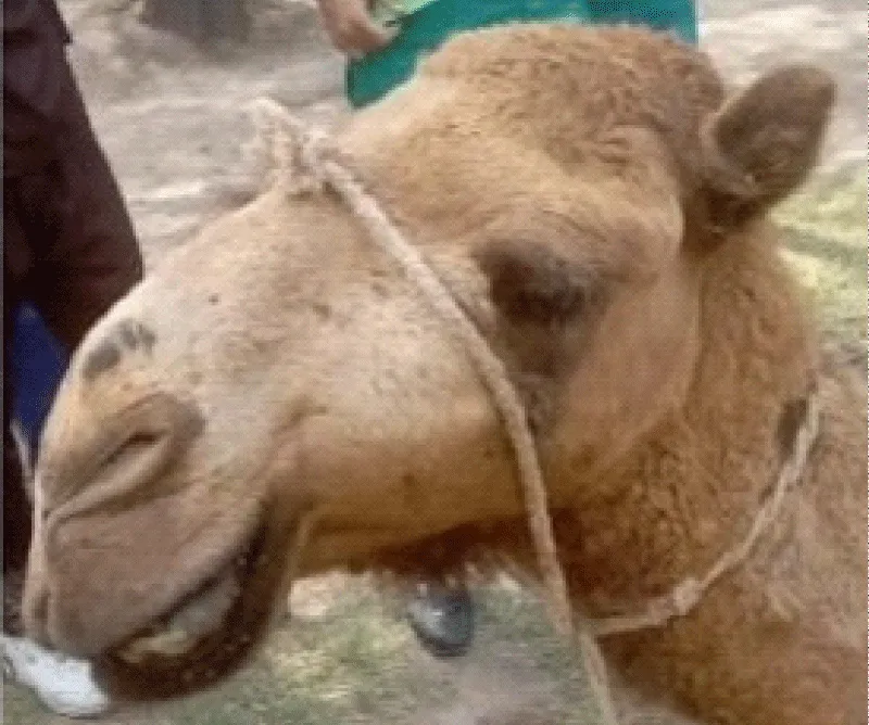
Figure 1: An MRI image showing an acute left paramedian thalamic infarction.
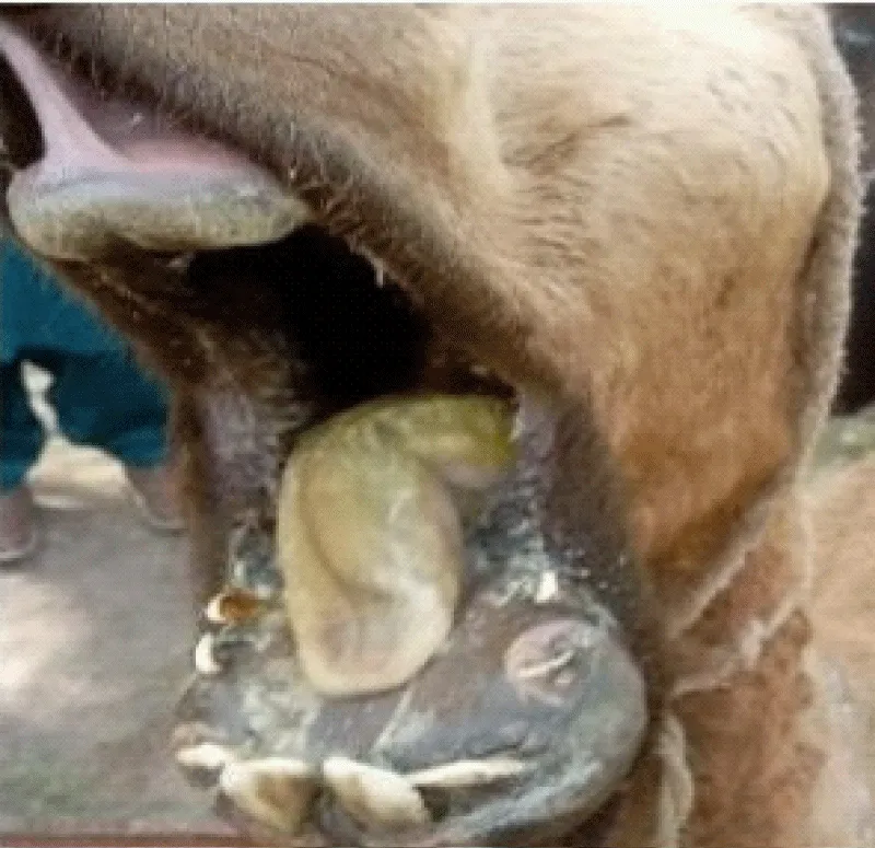
Figure 2: in oral cavity draining from upper Premolar tooth.
Physical examination, diagnosis, and treatment
Due to the narrow aperture of the camel oral cavity, it was difficult to thorough examination of the oral cavity [1,14-16]. The handlers restraining the animal enabled a physical examination of the oral cavity, which revealed chronic gingivitis around the upper premolar tooth root. This had led to sinus formation, with pus oozing from the sinus tract. Based on the history, physical findings, and clinical examination, the animal was diagnosed with a periapical tooth root infection. Treatment was then prescribed for a 4-week duration [1,2,6,17].
The following treatment protocol was instituted for four weeks. Conservative medical management using systemic antibiotics and anti-inflammatory drugs has been recommended for early or moderate cases of dental abscessation in large animals [5,6,16,18].
- Inj.Clavet. 1ml per 20 kg body weight by intramuscular route once daily for 3-5 days
- Inj.Ketoject 1ml per 33 kg body weight by intramuscular route once daily for 3-5 days
- Local application of boro glycerine gel twice daily
- Flushing of the oral cavity with normal saline thrice daily
Local antiseptic treatment and regular flushing of the oral cavity help reduce bacterial contamination and promote healing of infected periodontal tissues [5,6].
The animal was re-examined after one week and showed positive recovery. Long-term antibiotic therapy was continued for four weeks, after which the animal showed uneventful recovery. Similar therapeutic outcomes following conservative management of dental infections in camelids have been reported in previous studies [5,7]. The animal was re-examined after one week and showed positive results of recovery. The long-term use of antibiotics was continued until 4 weeks after that animal get an uneventful recovery.
Dental disorders in camels, particularly tooth root
abscesses and periodontal infections, are frequently reported in animals between four and eight years of age when permanent teeth are erupting [1,2,4,6]. During this period, the penetration of feed particles into the periodontal membrane may predispose animals to bacterial infections that subsequently develop into periapical abscesses. In many cases, these infections are associated with feeding on coarse or fibrous fodder that can cause trauma to the gingival tissues and promote microbial invasion [2,11].
The clinical signs observed in the present case, including facial swelling below the eye, reduced feed intake, and decreased milk production, are consistent with the typical manifestations of dental infections reported in camelids [4,7,11]. Facial swelling in the maxillary region often indicates involvement of the upper premolar or molar teeth, where infection may spread through surrounding tissues and result in sinus tract formation with purulent discharge. Such conditions can significantly affect the general health and productivity of the animal if not diagnosed and treated promptly.
Diagnosis of periapical tooth root infections in camels is usually based on a combination of clinical history, physical examination, and, when available, radiographic evaluation [1,7,14]. However, due to the anatomical structure and narrow oral cavity of camels, thorough oral examination can be challenging under field conditions. In the present case, diagnosis was primarily based on clinical findings, including gingivitis, sinus formation, and the presence of pus discharge from the affected tooth region.
The treatment approach used in this case involved systemic antibiotic therapy combined with anti-inflammatory medication and local oral management [5,6,18]. Administration of broad-spectrum antibiotics helps control bacterial infection, while anti-inflammatory drugs reduce pain and swelling, improving the animal’s ability to eat normally. In addition, local treatment with antiseptic gel and regular flushing of the oral cavity helps reduce bacterial load and promotes healing of infected tissues.
The positive response to treatment observed in this case indicates that conservative medical management can be effective when the infection is detected at an early stage and appropriate supportive care is provided. Continuous monitoring during the treatment period is essential to ensure resolution of infection and to prevent recurrence. Similar therapeutic approaches have been reported to yield successful outcomes in cases where surgical extraction of the affected tooth is not immediately required [1,4,7].
Overall, this case highlights the importance of early detection and timely veterinary intervention in managing dental infections in camels. Proper feeding management, regular oral examination, and prompt treatment of gingival inflammation can help reduce the risk of progression to severe tooth root infections.
Periapical tooth root infection is an important dental condition that can adversely affect the health, feeding behavior, and productivity of camels. The present case demonstrates that early diagnosis based on clinical examination, followed by appropriate systemic antibiotic therapy and local oral care, can lead to successful recovery without the need for surgical intervention. Regular monitoring of oral health and prompt management of gingival infections are essential to prevent the development of more severe dental complications. Increased awareness among camel owners and veterinarians regarding dental diseases can contribute to improved animal welfare and sustained productivity in camel farming systems.
Informed consent
Owner has orally provided consent for the clinical treatment and subsequent publication.
Author contributions
Ayat Fatima*Writing original manuscript, Sayyed Aun Muhammad, Hamid Akbar and abdul mateen has guided, drafted and review the original manuscript.
- Niehaus AJ, Anderson DE. Dental disorders in camelids: diagnosis and management. Vet Clin North Am Food Anim Pract. 2021;37(1):125-140.
- Van Amstel S. Dental diseases in camelids and their clinical significance. J Camel Pract Res. 2020;27(2):145-152.
- Cebra ML, Cebra CK, Garry FB. Tooth root abscesses in New World camelids: 23 cases. J Am Vet Med Assoc. 1996;209(4):819-822. Available from: https://pubmed.ncbi.nlm.nih.gov/8756887/
- Proost K, Pardon B, Pollaris E, Ducatelle R, Vlaminck L. Dental disease in alpacas: prevalence and clinical findings. J Vet Intern Med. 2020;34(2):847-856.
- Rahman MS, Ahmed S, Rahman MM. Management of dental abscesses in large animals: clinical perspectives. Vet Med Sci. 2023;9(5):2312-2321.
- Smith BB, Modransky P. Advances in camelid dentistry and oral health management. Animals (Basel). 2022;12(18):2441.
- Sparnon A, Smith J, Mulon PY, Hecht S, Anderson D, Van Amstel S. Tooth root abscess and mandibular infections in Old World camelids: three case reports. Case Rep Vet Med. 2024;2024:4589572.
- Ahmed A, Al-Swailem A, Al-Khalifa H. Clinical evaluation of oral and dental disorders in dromedary camels. Vet World. 2022;15(4):1032-1038.
- Hussein MF, Al-Dughaym AM. Camel diseases and veterinary management in arid regions. Front Vet Sci. 2021;8:642965.
- Niehaus AJ. Dental disease in llamas and alpacas. Vet Clin North Am Food Anim Pract. 2009;25(2):281-293. Available from: https://doi.org/10.1016/j.cvfa.2009.03.007
- Anderson DE, Whitehead CE. Diseases of the teeth and oral cavity in camelids. In: Smith BP, editor. Large Animal Internal Medicine. 5th ed. St Louis: Mosby; 2015. p. 1201-1210.
- Smith BB. Dental disease in camelids. In: Smith BP, editor. Large Animal Internal Medicine. St Louis: Mosby; 2006. p. 1189-1198.
- Van Amstel S, Shearer JK. Manual for Treatment and Control of Lameness in Cattle and Camelids. Ames: Wiley-Blackwell; 2006. Available from: https://rexresearch1.com/CattleLibrary/ManualTreatmentControlLamenessCattle.pdf
- Radostits OM, Gay CC, Hinchcliff KW, Constable PD. Veterinary Medicine: A Textbook of the Diseases of Cattle, Horses, Sheep, Pigs and Goats. 10th ed. London: Saunders Elsevier; 2007.
- Fowler ME. Medicine and Surgery of Camelids. 3rd ed. Ames: Wiley-Blackwell; 2010. Available from: https://www.wiley.com/en-dk/Medicine+and+Surgery+of+CamelidsC+3rd+Edition-p-9780470961698